




Abstract
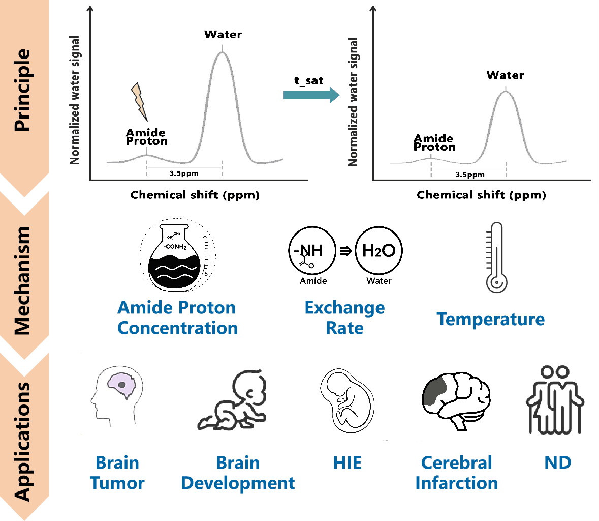
Amide proton transfer (APT) imaging, as a novel magnetic resonance molecular imaging technique based on the chemical exchange saturation transfer (CEST) effect, indirectly reflects molecular-level information such as tissue protein concentration, pH, and microenvironment by detecting the exchange between intracellular free water and amide protons in proteins/peptides. Since it was first proposed by Professor Jinyuan Zhou in 2003, the technique has achieved significant progress in theoretical modeling, sequence optimization, and clinical translation, particularly in its expanding application to central nervous system diseases. In recent years, APT has been widely used for tumor grading, stroke staging, early identification of hypoxic-ischemic encephalopathy (HIE), and pathological tracking of neurodegenerative diseases. This review systematically summarizes the principles and advantages of APT imaging, with a focus on its recent applications in brain tumors, cerebral infarction, and neurodegenerative diseases. It also discusses signal variation characteristics and underlying mechanisms in different disease states, as well as current clinical applications and technical challenges.
Keywords:Amide proton transfer (APT); chemical exchange saturation transfer (CEST); magnetic resonance molecular imaging; central nervous system (CNS); brain tumor; cerebral infarction; neurodegenerative disease
Introduction
Chemical exchange saturation transfer (CEST) magnetic resonance imaging is a molecular imaging technique based on the chemical exchange between exchangeable protons and water molecules. Through frequency-selective saturation and indirect detection, it can non-invasively detect various endogenous or exogenous metabolites containing exchangeable proton groups, and is widely used to obtain metabolic and microenvironmental information from tissues. Amide proton transfer (APT) imaging is one of the most clinically translated CEST techniques and was first reported by Zhou et al. in Nature Medicine in 2003[1] , drawing wide attention. Unlike conventional MRI, APT imaging does not require exogenous contrast agents and can acquire molecular-level information such as mobile protein/peptide content and pH environment within tissues, offering a new perspective for metabolic evaluation and disease mechanism research. Over the past two decades, APT has rapidly expanded from animal experiments [2] to human studies[3-5] , with increasing use in a variety of central nervous system (CNS) disorders including brain tumors, stroke, hypoxic-ischemic encephalopathy (HIE), and neurodegenerative diseases. While previous reviews have discussed APT imaging in individual disease entities, such as glioma or stroke, few have systematically examined shared mechanisms and signal characteristics across CNS pathologies.
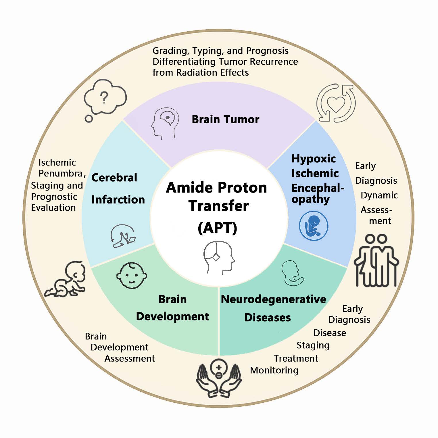
This review seeks to address a systematic overview of APT imaging, not only covering its principles and technical aspects, but also emphasizing disease-specific signal mechanisms and clinical applications across major CNS disorders. Particular attention is given to the shared and divergent mechanisms of APT contrast in tumors, ischemia, hypoxia, and proteinopathies, as well as the clinical implications for diagnosis, staging, and prognosis. Furthermore, we discuss current technical limitations and the challenges that must be addressed to facilitate broader clinical adoption of APT imaging. Through this comparative approach, we aim to provide readers with an integrative understanding of APT’s diagnostic and translational value in CNS disease (Figure 1 ).
Figure 1. Amide proton transfer imaging applications in central nervous system diseases.
Principles and Technical Characteristics of APT Imaging
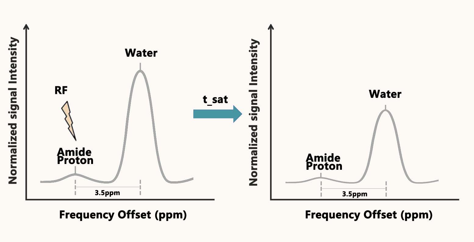
APT imaging is a molecular MRI technique based on the CEST mechanism. Its basic principle[1] involves applying a frequency-selective saturation radiofrequency pulse at approximately +3.5 ppm from the water resonance, which saturates the amide protons (–NH) within proteins and peptides in tissues. These saturated protons then exchange with water protons, leading to a decrease in water signal intensity. By comparing the water signal before and after saturation, one can indirectly assess the concentration of mobile proteins/peptides and local metabolic changes in tissues (Figure 2).
Figure 2. Schematic illustration of the principle of amide proton transfer (APT) imaging.
The APTw signal intensity depends not only on the concentration of exchangeable amide protons but also on their exchange rate, which is highly influenced by tissue pH and temperature[1] . Therefore, APT can reflect protein metabolism and is particularly sensitive to metabolic disturbances such as tissue acidosis. It complements techniques such as diffusion-weighted imaging (DWI), perfusion-weighted imaging (PWI), and magnetic resonance spectroscopy (MRS) in evaluating molecular metabolism, tissue acidity, and protein content, making it a valuable tool for studying various central nervous system diseases. APTw signal acquisition is influenced by multiple imaging parameters. Common settings include: radiofrequency saturation power B₁ = 1.5–2.5 μT, saturation duration t_sat = 0.8–2.0 s, and 25–61 offset frequency points for Z-spectrum acquisition. The APTw signal is usually quantified using the magnetization transfer ratio (MTR) asymmetry at 3.5 ppm, MTR_asym(3.5 ppm). Although this method is simple and practical, it is susceptible to interference from other CEST effects or non-exchangeable background signals, and its specificity and quantitative accuracy remain subjects of debate[6] .
To better quantify the APT effect, the magnetization transfer ratio asymmetry (MTRasym) is the most commonly used semi-quantitative index, defined as[7] :
MTRasym (Δω) = [Ssat(–Δω)–Ssat(+Δω)] / S₀
where Ssat (±Δω) represent the signal intensities at frequency offsets ±Δω from the water resonance (typically Δω = 3.5 ppm for APT), and S₀is the unsaturated signal. This asymmetry approach reflects the net saturation transfer from amide protons to water and serves as an indirect marker of protein content and exchange processes.
Advances in the Application of APT Imaging in Brain Tumors
Glioma Grading, Typing, and Prognosis
As a molecular imaging technique based on endogenous protein contrast, APT imaging has been widely used in recent years for grading and molecular subtyping of brain gliomas. Multiple studies have consistently reported a positive correlation between APTw signal intensity and glioma pathological grade, with high APTw signals commonly seen in high-grade gliomas. These hyperintense signals are often evident even in lesions lacking contrast enhancement[8] . Togao et al.[9] further reported that in diffuse gliomas without typical enhancement, APT imaging can compensate for the limitations of structural, DWI, and perfusion imaging by providing molecular contrast information beyond anatomical imaging, thus reflecting mobile protein/peptide concentrations and improving grading accuracy.
In addition to its grading value, APTw signal has also been explored as a marker for tumor biological behavior and patient prognosis. Almeida et al.[10] found that in WHO grade 4 gliomas, APT values were significantly associated with 1-year survival. Studies by Joo[7] and Paech[11] also indicated that elevated APTw signal was an independent predictor of poor prognosis. These findings suggest that APT can detect spatial heterogeneity in intratumoral metabolic activity and may serve as a noninvasive prognostic molecular imaging biomarker. However, most studies have used cross-sectional designs, and the causal relationship between APTw signal variation and survival outcomes remains unclear. Multicenter validation is required to confirm its predictive stability. With the 2016 and 2021 WHO classifications of central nervous system tumors incorporating molecular markers into diagnostic criteria, the potential of APT for preoperative molecular prediction has attracted increasing attention. APT values correlate with IDH mutation status, being lower in IDH-mutant than in IDH-wildtype gliomas,[12] likely reflecting underlying metabolic and proliferative differences, and they also display a negative association with Ki-67[13] .Multimodal radiomics models incorporating APT, DWI, and DCE imaging demonstrated high accuracy in predicting IDH-1 mutation status and Ki-67 expression. However, the ability of APT to distinguish O6-methylguanine-DNA methyltransferase (MGMT) promoter methylation status remains uncertain, as most studies report no significant signal differences[7,10] .
Differentiation of other Brain Tumors
Beyond gliomas, APT imaging has demonstrated diagnostic utility in differentiating other common intracranial tumors by providing molecular-level information that complements conventional MRI. Recent studies have explored its value in distinguishing solitary brain metastases (SBMs) from glioblastoma multiforme (GBM). Yu et al. demonstrated that APTw signal intensities were significantly higher in GBM than in SBMs in the peritumoral brain zone[15] , and Chen et al. also found that the APTw values and CBF in the peritumoral region were significantly higher in GBM compared to SBM[16] , likely reflecting the higher concentration of mobile proteins and peptides due to GBM’s more aggressive proliferative and infiltrative behavior. The APTw signal heterogeneity was also greater in GBM, offering an additional dimension for tumor characterization[15] . The combined application of APTw with other functional imaging parameters can significantly improve diagnostic accuracy[16,17] .
APT imaging has also been applied to characterize meningiomas, which represent another common extra-axial tumor. Joo et al. [18] investigated the value of amide proton transfer (APT) imaging in differentiating benign from atypical meningiomas. The results demonstrated that the MTRasym values were significantly higher in atypical meningiomas compared to benign ones, suggesting that APT imaging may serve as a useful adjunct to conventional MRI by enhancing the ability to distinguish atypical meningiomas. Koike et al.[19] conducted a pilot APT-CEST study in seventeen patients and found that lesions that later exhibited significant volumetric growth on follow-up exhibited markedly elevated mean MTR. Moreover, APTw signal intensity correlated linearly with the annual percentage increase in tumour diameter, whereas conventional MRI features offered no prognostic utility. These observations imply that the protein-weighted contrast of APT can unmask the latent proliferative drive of even WHO-grade I meningiomas, allowing clinicians to foresee which innocuous-looking masses may one day demand intervention. These findings support the utility of APT imaging in the noninvasive assessment of tumor biology across diverse brain tumor types.
Differentiating Tumor Recurrence from Radiation Effects
In post-surgical or post-radiotherapy follow-up of brain tumors, distinguishing tumor recurrence from radiation necrosis or other treatment-related effects remains a major imaging challenge. Conventional contrast-enhanced MRI has limitations, especially when enhancement patterns are atypical or overlap between entities occurs. Recently, APT imaging has been investigated as a tool for this differential diagnosis. Several studies[20-23] have applied APT to differentiate true glioma recurrence from treatment-related changes, showing that combining APT with DWI, perfusion, and other MRI techniques significantly improves diagnostic performance. For example, Chen et al.[24] reported that APT imaging alone achieved pooled sensitivity and specificity of 0.85 and 0.88, respectively, which increased to 0.92 and 0.83 when combined with additional MRI parameters. A very recent meta-analysis by Essed et al.[5] confirmed the diagnostic utility of APT in differentiating true progression from treatment effects, with reported sensitivity and specificity of 0.88 and 0.84.
In addition, Jiang et al.[25] demonstrated that biopsy guidance based on APT imaging improves sampling accuracy. High-signal APT regions typically correspond to viable tumor tissue, while low-signal areas are often necrotic. These findings support the use of APT as a noninvasive molecular imaging tool to aid in the identification of indeterminate postoperative lesions and to guide subsequent treatment strategies.
Advances in the Application of APT Imaging in Cerebral Infarction
As early as 2003, Professor Jinyuan Zhou, the inventor of APT, demonstrated using a rat model that APT could detect pH changes in ischemic brain regions.[1] Subsequent animal and clinical studies have further explored the application value of APT in stroke.
APT Imaging of Acute Infarction and Ischemic Penumbra
Animal experiments[26] showed that within 0.5–3 hours after middle cerebral artery occlusion (MCAO), the APTw signal significantly decreased in the ischemic region and correlates closely with areas showing diffusion restriction, even when T1WI and T2WI remain normal or show only mild changes. This suggests that APT may outperform structural imaging in identifying early metabolic abnormalities. In a clinical study, Heo et al.[27] observed that in ischemic penumbra regions with hypoperfusion but no irreversible injury, APTw signal was reduced due to pH drop from restricted metabolism. Conversely, in “benign oligemia” areas with hypoperfusion but preserved pH, APTw signals remained near normal. Thus, APT may assist in differentiating reversible from irreversible ischemic tissue and help delineate the penumbra more accurately. Further studies found that APTw signal reduction areas were typically larger than diffusion-positive regions (DWI high signal) but smaller than perfusion-deficient zones (PWI), and the mismatch between APT and DWI regions may serve as an additional indicator of the ischemic penumbra[28] . This unique imaging pattern provides supplementary information beyond the classic perfusion–diffusion mismatch and may refine patient selection for thrombolytic or thrombectomy treatment in the hyperacute stage.
Infarct Staging and Prognostic Evaluation
During the subacute to chronic phases of cerebral infarction, ongoing changes in tissue metabolism and microenvironmental pH are crucial for assessing disease progression and prognosis. Due to its pH sensitivity, APT imaging has been proposed as a tool for dynamic monitoring of post-stroke metabolic recovery. Several studies have evaluated its utility in infarct staging and prognostic stratification. For example, Song et al.[29] analyzed APT images in patients within 2 hours to 7 days of stroke onset and found that APTw signal in the ischemic region remained lower than that in contralateral white matter, with a time-dependent upward trend, likely reflecting gradual resolution of acidosis. Momosaka et al.[30] further showed that multipercentile APT parameters significantly correlated with clinical outcomes, suggesting that APT may quantify the severity of metabolic dysfunction and aid in prognostic stratification.
It is noteworthy that APT alone has limited predictive power. A multimodal imaging study by Zhou et al.[31] demonstrated that the minimum APT value showed the strongest correlation with NIHSS scores, but only in combination with apparent diffusion coefficient (ADC) and computed tomography perfusion (CTP) did the prognostic model achieve optimal performance. This supports the view that APT complements rather than replaces existing imaging markers. Similarly, studies by Zhang et al.[32] and Yu et al.[33] , focusing on reperfusion efficacy and clinical recovery, found that rising APTw signal trends aligned with symptomatic improvement, further reinforcing its role in disease monitoring.
Application of APT Imaging in Hypoxic-Ischemic Encephalopathy (HIE)
Neonatal hypoxic-ischemic encephalopathy (HIE) is a major cause of severe brain injury in newborns. Timely detection of metabolic abnormalities is crucial for prognosis assessment. In animal experiments, Zheng et al.[34] established a neonatal piglet model of hypoxic-ischemic brain injury (HIBI) and employed amide proton transfer (APT) imaging combined with proton magnetic resonance spectroscopy (¹H-MRS) to evaluate cerebral metabolic changes. The results demonstrated a significant decrease in APTw signal intensity and a marked elevation in lactate levels in the basal ganglia following hypoxic-ischemic insult, indicating local tissue acidosis and lactate accumulation. The reduction in APTw signal was primarily attributed to the decreased exchange rate of amide protons with water in an acidic environment. Notably, follow-up observations revealed that the recovery of APTw signal preceded the decline in lactate concentration, suggesting that APT imaging may serve as a sensitive tool for dynamically monitoring acid-base metabolic recovery and thus assessing the extent of tissue injury and potential prognosis. In human studies, Chen et al.[35] reported that APT imaging may facilitate the early identification of mild hypoxic-ischemic encephalopathy (HIE) cases that appear negative on conventional MRI. Other studies have shown increased APTw signal intensity in multiple brain regions of infants with HIE, with APT demonstrating higher sensitivity for HIE diagnosis compared to ADC measurements[36,37] . These findings suggest that APT imaging holds promise as an early diagnostic tool for HIE.
Application of APT Imaging in Pediatric Brain Development
Myelination of white matter during childhood is a critical process in the development of the nervous system. Conventional MRI can assess myelination using T1- and T2-weighted imaging, but this is primarily morphological and lacks molecular-level quantitative indicators. The combination of MTR and APT techniques offers complementary molecular imaging approaches for evaluating the microstructural development of white matter[38] .
Zheng et al.[39] applied APT and MTR imaging to assess brain development in 38 neonates without neurological disease and found that APT and MTR values in the frontal lobe, basal ganglia, and occipital lobe were positively correlated with gestational age, suggesting the potential of these metrics for reflecting white matter maturity. Tang et al.[38] further investigated white matter characteristics in children with developmental delay and found that in the group with delayed myelination, MTR values were generally lower across all brain regions, indicating reduced myelin lipid synthesis; meanwhile, APT values were significantly elevated in regions such as the pons, middle cerebellar peduncles, corpus callosum, frontal and occipital white matter, and centrum semiovale, possibly due to a relatively higher proportion of protein content around unmyelinated axons. In contrast, children with developmental delay but normal myelination showed no significant differences in APT or MTR values compared to healthy controls. APT imaging thus provides a novel method for the early quantitative assessment of white matter development. Particularly in cases where conventional MRI indicates delayed myelination, APT and MTR offer biologically meaningful parameters that may aid in the early identification of developmental delay. In addition, APT imaging also holds significant value for studying the developmental trajectory of normal pediatric brains. Literature reports[40] indicate that APT values in white matter exhibit a downward trend with increasing age, with the most pronounced changes occurring during the first year after birth, reflecting the rapid progression of the myelination process. This change is particularly evident in the corpus callosum, frontal and occipital white matter, and the centrum semiovale, suggesting that APT imaging is highly sensitive to dynamic changes in white matter maturation.
Application of APT Imaging in Neurodegenerative Diseases
Neurodegenerative diseases, such as Alzheimer’s disease (AD), mild cognitive impairment (MCI), and Parkinson’s disease (PD), are typically characterized by neuronal loss, abnormal protein aggregation, and glial activation in specific brain regions. These pathological changes may alter the local protein content within brain tissue, thereby affecting APT imaging signals and offering a new, noninvasive approach for exploring disease mechanisms and facilitating early diagnosis.
Application of APT Imaging in AD and MCI
The core pathological features of AD include excessive deposition of β-amyloid plaques and tau-related neurofibrillary tangles in the brain, accompanied by widespread neuronal and synaptic loss as well as glial activation[41] . These changes result in abnormal protein metabolism within brain tissue, which in turn affects APT imaging signals.
Wang et al.[42] performed APT imaging on AD patients using a 3.0T MRI system and found that APT values in the bilateral hippocampus were significantly higher than those in age-matched healthy controls. The elevated APTw signal may reflect increased levels of mobile proteins due to abnormal protein aggregation or glial proliferation. Moreover, the study showed a significant negative correlation between hippocampal APT values and Mini-Mental State Examination (MMSE) scores, suggesting that APT imaging may serve as a potential indicator of disease severity. In a separate study, Wang et al.[43] observed decreased APT values in the hippocampus and cortex of AD model rats, which correlated with histological findings of neuronal loss and increased GFAP-positive astrocytes, indicating a close relationship between APTw signal alterations and underlying tissue changes. Subsequent studies in AD and MCI populations have confirmed similar trends: APTw signal intensity in specific brain regions is higher in patients with AD[44] and MCI[45,46] compared to healthy elderly controls. Guo et al.[46] observed significantly increased APT values in the bilateral hippocampus and amygdala in 70 patients with amnestic MCI (aMCI). The APT value of the left amygdala yielded an AUC of 0.875 for diagnosing aMCI. In a study by Chen et al.[47] , combining the APT value of the right hippocampus with the level of glutamate–glutamine (Glx) significantly improved the diagnostic performance for aMCI compared to either parameter alone. These findings suggest that APT imaging has favorable diagnostic performance in the aMCI population and holds promise as a novel, noninvasive molecular imaging biomarker for aMCI.
Application of APT Imaging in PD
The primary pathological features of PD include progressive loss of dopaminergic neurons in the substantia nigra and formation of Lewy bodies (aggregates of α-synuclein). In addition to the substantia nigra, PD may also involve widespread metabolic changes in cortical regions, which can be reflected in APT imaging alterations.
Li et al.[48] conducted a study using CEST imaging in 61 PD patients and 24 healthy controls, and found that APTw signal and total CEST signal in the substantia nigra decreased significantly with disease progression. These decreases were negatively correlated with Hoehn and Yahr (H&Y) staging, Unified Parkinson’s Disease Rating Scale (UPDRS) scores, and disease duration. The changes in APTw signal may be attributed to reduced concentrations of mobile proteins and peptides due to dopaminergic neuronal loss. In PD patients with asymmetric motor symptoms, the substantia nigra on the more affected side exhibited lower APTw signal, and the calculated asymmetry index was positively correlated with clinical motor symptom asymmetry[49] . These findings indicate that APT not only quantifies pathological changes in the substantia nigra, but also detects lateralized differences that correlate with clinical symptom asymmetry, suggesting potential utility in PD imaging subtyping and lateralized treatment planning. Beyond the substantia nigra, PD may also affect the cerebral cortex. Tian et al.[50] applied 3D-APT imaging in 34 PD patients and performed whole-brain analysis, revealing increased APT values in multiple regions including the temporal cortex. Although no significant correlations were found between APTw signal and disease duration or clinical scores, the APT parameter achieved an AUC of 0.865 for differentiating PD from controls, and the AUC increased to 0.932 when combined with multiparametric features. This study suggests that APT abnormalities are not limited to deep nuclei and may reflect widespread synucleinopathy-related metabolic alterations in the cortex. Collectively, the studies by Li[48] and Tian[50] suggest that APTw signal changes in PD may exhibit region-specific patterns.
Discussion
Mechanistic Characteristics of APT Imaging in Central Nervous System Diseases
APT imaging is a molecular MRI technique sensitive to the exchange of amide protons between mobile proteins/peptides and water. This exchange mainly depends on the concentration of mobile proteins/peptides, local tissue pH, and temperature. However, as body temperature remains stable in humans, protein concentration and pH are generally considered the primary determinants of APT signal variation in clinical imaging. Additional factors, such as tissue water content and magnetization transfer effects, can also influence APT signal intensity to a lesser extent[1] . By sensitively reflecting these properties, APT provides a novel molecular perspective on CNS pathology ( table 1 ), complementing conventional MRI. Below, we discuss the mechanistic contributors to APT signal alterations across CNS diseases, organized by the underlying biochemical factors that drive these changes.
Table 1. Summary of APT Signal Characteristics and Mechanisms in Major CNS Diseases
|
Disease Type |
APT Signal Change |
Primary Mechanism |
Clinical Utility |
|
High-grade glioma |
Increased |
Increased protein content, active cellular proliferation |
Grading, molecular subtyping, recurrence monitoring |
|
Acute cerebral infarction |
Decreased |
Tissue acidosis, decreased pH |
Early detection, penumbra localization |
|
Neonatal HIE |
Increased / Decreased |
pH alteration / abnormal protein or metabolic accumulation |
Early diagnosis in MRI-negative cases |
|
Developmental delay |
Increased |
Delayed myelination, increased intracellular mobile protein ratio |
Quantitative assessment of myelination |
|
Alzheimer’s disease |
Increased |
Abnormal protein aggregation, neuroinflammation, neuronal loss |
Early detection, biomarker exploration |
|
Parkinson’s disease |
Decreased (Substantia nigra) Increased(cortex) |
Substantia nigra neuronal loss/ synuclein aggregation |
Disease staging, monitoring, Assessment of cortical alterations |
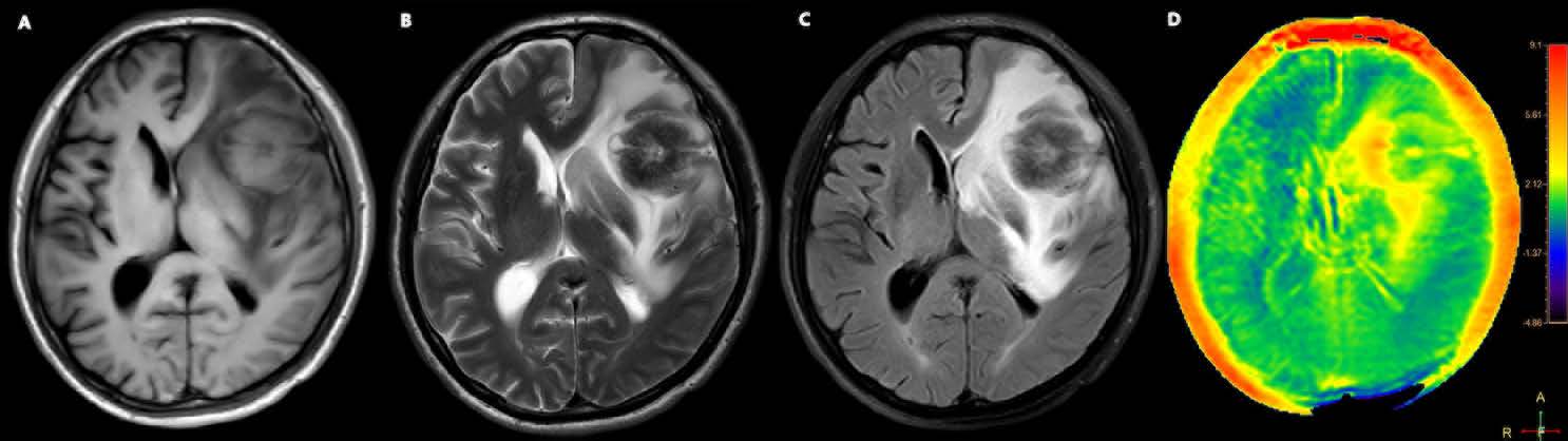
The APTw signal intensity directly correlates with mobile protein and peptide concentration. Elevated APT signals often indicate increased mobile protein content in tissues. In brain tumors (Figure 3 and Figure 4), rapid cellular proliferation leads to a high concentration of cytosolic proteins/peptides, which increases the APTw signal. This mechanistic link explains why APT hyperintensity correlates with more aggressive tumor activity and has shown utility in glioma grading and molecular subtype prediction.[10] Similarly, recurrent tumor and treatment-related necrosis can be distinguished by APT. Recurrent tumors tend to be APT-hyperintense due to viable tumor cells with high protein metabolism, whereas radiation necrosis shows low APT signal because the tissue is largely non-neoplastic, fibrotic or necrotic with minimal protein exchange[20,22] . The lack of active protein synthesis and metabolic activity in necrotic tissue results in fewer mobile amide protons and thus a diminished APT signal. This contrast in APT behavior provides a mechanistic basis for using APT imaging to differentiate residual/recurrent tumor from post-therapy necrosis in neuro-oncology.
Figure 3. A 71-year-old male with a solitary brain metastasis. The tumor was located in the left frontal lobe, demonstrating a slightly hypointense signal on T1WI (A), and a heterogeneously isointense to hypointense signal on both T2WI (B) and T2-FLAIR (C) sequences. The APTw image (D) shows a relatively homogeneous isointense mass.
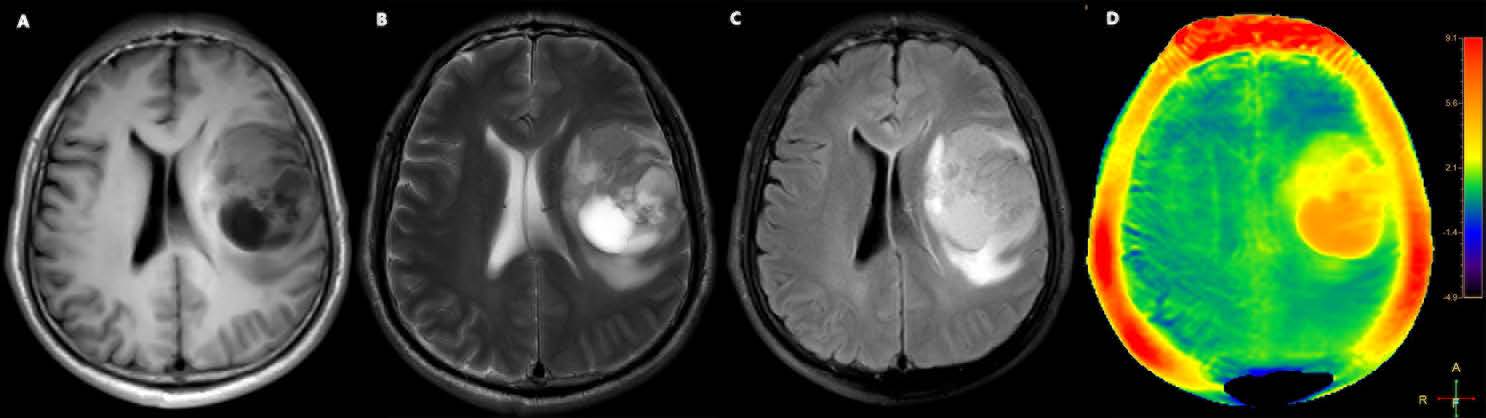
Figure 4. A 55-year-old male with glioblastoma. The tumor was located in the left frontal lobe, demonstrating a slightly hypointense signal on T1WI (A), and a heterogeneously isointense to hyperintense signal on both T2WI (B) and T2-FLAIR (C) sequences with cystic components. The APTw image (D) shows a heterogeneously hyperintense mass.
Beyond tumors, developmental and neurodegenerative contexts also highlight the role of protein content. In the developing pediatric brain, regions of delayed myelination or immature white matter show higher APT signals due to a greater pool of mobile cytosolic proteins, as myelin formation, tracked by magnetization transfer imaging, gradually sequesters lipids and proteins into solid myelin structures[51] . Studies have demonstrated that APT imaging can sensitively monitor myelination processes and white matter maturation, offering molecular imaging support for tracking white matter injury and recovery in preterm infants and other neurodevelopmental disorders[38,39] . Likewise, abnormally high APTw signals observed in neurodegenerative diseases such as Alzheimer’s disease might reflect protein accumulation or redistribution. For instance, excessive amounts of misfolded or aggregated proteins and reactive glial proteins in affected brain regions, though the mobile fraction of these proteins and their exact contribution to APT remain to be clarified. However, immobile fibrillar protein aggregates like amyloid plaques do not directly increase APT signals; rather, the surrounding microenvironmental changes involving mobile proteins from degenerating neurons or activated glia primarily contribute to these APT elevations. An increase in mobile protein/peptide concentration generally drives APT signal up, serving as an indicator of active protein metabolism or accumulation in tissue.
Intracellular and extracellular pH strongly influences APT signals by affecting the proton exchange rate. Tissue acidosis (low pH) slows this exchange, reducing APT signals. Thus, APT imaging is highly sensitive to metabolic disturbances that alter pH. In acute cerebral infarction, impaired blood flow triggers anaerobic metabolism, lactic acid accumulation, and tissue acidosis, significantly decreasing APT signals shortly after stroke onset[30] . APT can therefore detect ultra-early ischemic changes, potentially before irreversible injury occurs. However, distinguishing reversible ischemic tissue (penumbra) from irreversibly damaged regions using APT alone remains uncertain. Interpretation typically requires complementary MRI methods like DWI and PWI, combined with clinical outcomes. Current stroke studies, limited by small sample sizes, still require histopathological validation to confirm whether APT reliably identifies metabolically compromised yet viable tissues.
Similarly, HIE involves metabolic acidosis following hypoxia. Preliminary studies indicate APT imaging sensitively detects early metabolic disruptions in neonatal brains, suggesting potential for early diagnosis and therapy monitoring[36,37] . However, further validation from large-scale studies remains necessary. Chronic neuroinflammation in neurodegenerative conditions may subtly alter local pH, indirectly influencing APT signals alongside protein changes. This pH-dependent characteristic underscores APT’s value in assessing ischemia and hypoxia-related disorders. Nonetheless, accurate clinical interpretation generally requires multimodal imaging approaches.
APT imaging primarily reflects protein concentration and pH. However, these signal changes lack disease specificity and require integrated clinical and imaging interpretation. Future research must validate APT signals through direct pathological and biochemical correlations. Integrating APT with complementary modalities such as MRS and PET can further enhance diagnostic accuracy, making APT a quantifiable and mechanism-driven clinical tool.
Methodological Advantages and Limitations
Compared to other metabolic imaging techniques such as MRS, which suffers from low spatial resolution and long scan times, and PET, which involves ionizing radiation and requires radiotracers, APT is more accessible and safer. It avoids exogenous agents and radiation exposure, while remaining sensitive to endogenous biomarkers like mobile proteins and pH.
However, several technical bottlenecks and challenges remain to be addressed before broader clinical application can be achieved.
First, APT imaging often suffers from low signal-to-noise ratio (SNR) , limited contrast, and high sensitivity to magnetic field inhomogeneities. The APT effect is highly susceptible to B0/B1 field inhomogeneity and noise interference, which can distort quantification and reduce diagnostic reliability[52] . This necessitates optimization of imaging parameters—such as increasing B₁ power, adjusting saturation duration, and adopting three-dimensional (3D) acquisition sequences—to enhance image stability and robustness. Despite ongoing efforts to correct for B0 and B1 inhomogeneities, accurate mapping and correction remain technically demanding, particularly in deep brain regions and heterogeneous field environments.
Second, the APT effect shows temperature dependence, which may affect interpretation in conditions with variable tissue temperature such as ischemia, fever, or intraoperative monitoring. Although generally stable in clinical settings, this factor should be considered in experimental designs and multicenter studies.
Third, specificity remains a major issue. The APTw signal arises not only from amide protons but is also influenced by other CEST effects. Overlapping contributions from various exchangeable components may cancel out or interfere with each other, thereby confounding the interpretation of true APTw signals[53] . To address this limitation, advanced post-processing techniques such as Lorentzian line-shape fitting and multi-pool modeling based on Bloch–McConnell equations have been proposed. Lorentzian fitting allows the separation of amide, nuclear Overhauser enhancement (NOE), and semi-solid magnetization transfer components within the Z-spectrum, while multi-pool models enable quantitative estimation of exchange parameters such as ksw, T1, T2, and proton concentrations. These approaches enhance the physiological interpretability of APTw signals and reduce contamination from non-specific background effects. However, their complexity, increased computational demands, and lack of standardization currently hinder widespread clinical adoption[54] .
Fourth, APT imaging is susceptible to partial volume effects, particularly in small anatomical structures such as brainstem nuclei, hippocampal subfields, or cortical layers. These effects may attenuate or mask pathological APTw signals, thereby complicating diagnostic interpretation and reducing sensitivity in small or heterogeneous regions.
Fifth, current studies have primarily focused on trends in APTw signal changes across disease states, while systematic validation of its diagnostic specificity remains lacking. Elevated APTw signal may occur in diverse pathological processes—such as tumor proliferation, gliosis, and synaptic alterations—making it difficult to determine etiology based solely on APT values. Furthermore, few studies have quantitatively correlated APT metrics with histopathological features in a systematic manner. Future research should incorporate multimodal imaging techniques such as MRS and PET, along with histological analysis to clarify the pathological basis of APTw signal alterations.
Finally, standardization and consistency must be improved. Substantial variability exists across studies in APT sequence implementation and data processing methods. This not only hampers comparability in multicenter studies but also restricts the broader clinical adoption of APT imaging. Moreover, APT imaging cannot independently provide sufficient diagnostic information and should be interpreted in conjunction with conventional MRI sequences and other functional imaging techniques to achieve comprehensive clinical insights. Although several consensus recommendations[6] for CEST and APT imaging have been proposed in recent years, further refinement and broader implementation of standardized acquisition protocols and quantification strategies—including emerging model-based methods—remain essential to ensure reproducibility and promote clinical translation.
Future Perspectives
Looking ahead, APT imaging holds promise for development in multiple directions within the field of central nervous system imaging. Multimodal integration is a key trend: combining APT with other imaging modalities such as DWI, arterial spin labeling (ASL), and PET—augmented by machine learning, radiomics, or image fusion techniques—may help unlock the full potential of APT imaging. This approach is expected to improve diagnostic accuracy and enable more comprehensive characterization of neurological diseases.
Conclusion
APT imaging has shown the greatest translational potential in tumor classification and grading, identification of ischemic penumbra in cerebral infarction and early diagnosis of HIE. Dynamic monitoring of APT values can also be applied for treatment response and prognosis evaluation. As APT transitions to clinical practice, resolving technical bottlenecks and fostering interdisciplinary collaboration, APT holds promise for contributing deeper insights into the precise diagnosis, therapeutic assessment, and pathophysiological understanding of brain disorders, thereby advancing the field of imaging medicine toward the molecular level.
Declarations
Author contributions
CY and YWM conceptualized the review and supervised the project. WWW conducted the literature review and drafted the manuscript. WWW, HJJ, and NW prepared the figures.
Acknowledgements
Not applicable.
Ethics approval and consent to participate
Not applicable.
Funding information
Not applicable.
Competing Interests
The authors declare that they have no existing or potential commercial or financial relationships that could create a conflict of interest at the time of conducting this study.
Data Availability
All data needed to evaluate the conclusions in the paper are present in the paper or the Supplementary Materials. Additional data related to this paper may be requested from the authors.
References
[1] Zhou J, Payen J, Wilson D, Traystman R, van Zijl P. (2003). Using the amide proton signals of intracellular proteins and peptides to detect pH effects in MRI. Nature medicine. 2003;9(8):1085-90. https://doi.org/10.1038/nm907
[2] Zhou J, Lal B, Wilson DA, Laterra J, van Zijl PC. (2003). Amide proton transfer (APT) contrast for imaging of brain tumors. Magnetic resonance in medicine. 2003;50(6):1120-6. https://doi.org/10.1002/mrm.10651
[3] Kulanthaivelu K, Jabeen S, Saini J, Raju S, Nalini A, Sadashiva N, et al. (2021). Amide proton transfer imaging for differentiation of tuberculomas from high-grade gliomas: Preliminary experience. The neuroradiology journal. 2021;34(5):440-8. https://doi.org/10.1177/19714009211002766
[4] Mu R, Qin X, Zheng W, Yang P, Huang B, Li X, et al. (2023). Amide proton transfer could be a surrogate imaging marker for predicting vascular cognitive impairment. Brain research bulletin. 2023;204:110793. https://doi.org/10.1016/j.brainresbull.2023.110793
[5] Essed RA, Prysiazhniuk Y, Wamelink IJ, Azizova A, Keil VC. (2025). Performance of amide proton transfer imaging to differentiate true progression from therapy-related changes in gliomas and metastases. European radiology. 2025;35(2):580-91. https://doi.org/10.1007/s00330-024-11004-y
[6] Zhou J, Zaiss M, Knutsson L, Sun PZ, Ahn SS, Aime S, et al. (2022). Review and consensus recommendations on clinical APT-weighted imaging approaches at 3T: Application to brain tumors. Magnetic resonance in medicine. 2022;88(2):546-74. https://doi.org/10.1002/mrm.29241
[7] Joo B, Han K, Ahn SS, Choi YS, Chang JH, Kang SG, et al. (2019). Amide proton transfer imaging might predict survival and IDH mutation status in high-grade glioma. European radiology. 2019;29(12):6643-52. https://doi.org/10.1007/s00330-019-06203-x
[8] Zhou J, Zhu H, Lim M, Blair L, Quinones-Hinojosa A, Messina SA, et al. (2013). Three-dimensional amide proton transfer MR imaging of gliomas: Initial experience and comparison with gadolinium enhancement. Journal of magnetic resonance imaging : JMRI. 2013;38(5):1119-28. https://doi.org/10.1002/jmri.24067
[9] Togao O, Hiwatashi A, Yamashita K, Kikuchi K, Keupp J, Yoshimoto K, et al. (2017). Grading diffuse gliomas without intense contrast enhancement by amide proton transfer MR imaging: comparisons with diffusion- and perfusion-weighted imaging. European radiology. 2017;27(2):578-88. https://doi.org/10.1007/s00330-016-4328-0
[10] Borges de Almeida G, Pascuzzo R, Mambrin F, Aquino D, Verri M, Moscatelli M, et al. (2024). The Role of Amide Proton Transfer (APT)-Weighted Imaging in Glioma: Assessment of Tumor Grading, Molecular Profile and Survival in Different Tumor Components. Cancers. 2024;16(17). https://doi.org/10.3390/cancers16173014
[11] Paech D, Dreher C, Regnery S, Meissner JE, Goerke S, Windschuh J, et al. (2019). Relaxation-compensated amide proton transfer (APT) MRI signal intensity is associated with survival and progression in high-grade glioma patients. European radiology. 2019;29(9):4957-67. https://doi.org/10.1007/s00330-019-06066-2
[12] Wu M, Jiang T, Guo M, Duan Y, Zhuo Z, Weng J, et al. (2024). Amide proton transfer-weighted imaging and derived radiomics in the classification of adult-type diffuse gliomas. European radiology. 2024;34(5):2986-96. https://doi.org/10.1007/s00330-023-10343-6
[13] Liang HX, Wang ZY, Li Y, Ren AN, Chen ZF, Wang XZ, et al. (2024). The application value of support vector machine model based on multimodal MRI in predicting IDH-1mutation and Ki-67 expression in glioma. BMC Med Imaging. 2024;24(1):244. https://doi.org/10.1186/s12880-024-01414-1
[14] Koike H, Morikawa M, Ishimaru H, Ideguchi R, Uetani M, Hiu T, et al. (2022). Amide proton transfer MRI differentiates between progressive multifocal leukoencephalopathy and malignant brain tumors: a pilot study. BMC Med Imaging. 2022;22(1):227. https://doi.org/10.1186/s12880-022-00959-3
[15] Yu H, Lou H, Zou T, Wang X, Jiang S, Huang Z, et al. (2017). Applying protein-based amide proton transfer MR imaging to distinguish solitary brain metastases from glioblastoma. European radiology. 2017;27(11):4516-24. https://doi.org/10.1007/s00330-017-4867-z
[16] Chen L, Li T, Li Y, Zhang J, Li S, Zhu L, et al. (2023). Combining amide proton transfer-weighted and arterial spin labeling imaging to differentiate solitary brain metastases from glioblastomas. Magnetic resonance imaging. 2023;102:96-102. https://doi.org/10.1016/j.mri.2023.05.004
[17] Knutsson M, Salomonsson T, Durmo F, Johansson ER, Seidemo A, Lätt J, et al. (2025). Differentiation between glioblastoma and solitary brain metastases using perfusion and amide proton transfer weighted MRI. Front Neurosci. 2025;19:1533799. https://doi.org/10.3389/fnins.2025.1533799
[18] Joo B, Han K, Choi YS, Lee SK, Ahn SS, Chang JH, et al. (2018). Amide proton transfer imaging for differentiation of benign and atypical meningiomas. European radiology. 2018;28(1):331-9. https://doi.org/10.1007/s00330-017-4962-1
[19] Koike H, Morikawa M, Ideguchi R, Uetani M, Hiu T, Matsuo T. (2022). Amide proton transfer and chemical exchange saturation transfer MRI differentiates between growing and non-growing intracranial meningiomas: a pilot study. Clinical radiology. 2022;77(4):e295-e301. https://doi.org/10.1016/j.crad.2021.12.017
[20] Huang Q, Wu J, Le N, Shen Y, Guo P, Schreck KC, et al. (2023). CEST2022: Amide proton transfer-weighted MRI improves the diagnostic performance of multiparametric non-contrast-enhanced MRI techniques in patients with post-treatment high-grade gliomas. Magnetic resonance imaging. 2023;102:222-8. https://doi.org/10.1016/j.mri.2023.06.003
[21] Jiang S, Guo P, Heo HY, Zhang Y, Wu J, Jin Y, et al. (2023). Radiomics analysis of amide proton transfer-weighted and structural MR images for treatment response assessment in malignant gliomas. NMR in biomedicine. 2023;36(1):e4824. https://doi.org/10.1002/nbm.4824
[22] Hou H, Diao Y, Yu J, Xu M, Wang L, Li Z, et al. (2023). Differentiation of true progression from treatment response in high-grade glioma treated with chemoradiation: a comparison study of 3D-APTW and 3D-PcASL imaging and DWI. NMR in biomedicine. 2023;36(1):e4821. https://doi.org/10.1002/nbm.4821
[23] Guo P, Unberath M, Heo HY, Eberhart CG, Lim M, Blakeley JO, et al. (2022). Learning-based analysis of amide proton transfer-weighted MRI to identify true progression in glioma patients. NeuroImage Clinical. 2022;35:103121. https://doi.org/10.1016/j.nicl.2022.103121
[24] Chen K, Jiang XW, Deng LJ, She HL. (2022). Differentiation between glioma recurrence and treatment effects using amide proton transfer imaging: A mini-Bayesian bivariate meta-analysis. Front Oncol. 2022;12:852076. https://doi.org/10.3389/fonc.2022.852076
[25] Jiang S, Eberhart CG, Zhang Y, Heo HY, Wen Z, Blair L, et al. (2017). Amide proton transfer-weighted magnetic resonance image-guided stereotactic biopsy in patients with newly diagnosed gliomas. Eur J Cancer. 2017;83:9-18. https://doi.org/10.1016/j.ejca.2017.06.009
[26] Jin T, Wang J, Chung J, Hitchens TK, Sun D, Mettenburg J, et al. (2024). Amide proton transfer MRI at 9.4 T for differentiating tissue acidosis in a rodent model of ischemic stroke. Magnetic resonance in medicine. 2024;92(5):2140-8. https://doi.org/10.1002/mrm.30194
[27] Heo HY, Zhang Y, Burton TM, Jiang S, Zhao Y, van Zijl PCM, et al. (2017). Improving the detection sensitivity of pH-weighted amide proton transfer MRI in acute stroke patients using extrapolated semisolid magnetization transfer reference signals. Magnetic resonance in medicine. 2017;78(3):871-80. https://doi.org/10.1002/mrm.26799
[28] Heo HY, Tee YK, Harston G, Leigh R, Chappell MA. (2023). Amide proton transfer imaging in stroke. NMR in biomedicine. 2023;36(6):e4734. https://doi.org/10.1002/nbm.4734
[29] Song G, Li C, Luo X, Zhao X, Zhang S, Zhang Y, et al. (2017). Evolution of Cerebral Ischemia Assessed by Amide Proton Transfer-Weighted MRI. Front Neurol. 2017;8:67. https://doi.org/10.3389/fneur.2017.00067
[30] Momosaka D, Togao O, Kikuchi K, Kikuchi Y, Wakisaka Y, Hiwatashi A. (2020). Correlations of amide proton transfer-weighted MRI of cerebral infarction with clinico-radiological findings. PloS one. 2020;15(8):e0237358. https://doi.org/10.1371/journal.pone.0237358
[31] Zhou L, Pan W, Huang R, Wang T, Wei Z, Wang H, et al. (2024). Amide Proton Transfer-Weighted MRI, Associations with Clinical Severity and Prognosis in Ischemic Strokes. Journal of magnetic resonance imaging : JMRI. 2024;60(6):2509-17. https://doi.org/10.1002/jmri.29333
[32] Zhang C, Yong X, Cao Y, Hsu YC, Shi H, Wu F, et al. (2025). Amide proton transfer MRI may reflect effective reperfusion and predict functional outcomes in patients with ischemic stroke. Journal of cerebral blood flow and metabolism : official journal of the International Society of Cerebral Blood Flow and Metabolism. 2025;45(3):421-30. https://doi.org/10.1177/0271678x241297110
[33] Yu L, Chen Y, Chen M, Luo X, Jiang S, Zhang Y, et al. (2019). Amide Proton Transfer MRI Signal as a Surrogate Biomarker of Ischemic Stroke Recovery in Patients With Supportive Treatment. Front Neurol. 2019;10:104. https://doi.org/10.3389/fneur.2019.00104
[34] Zheng Y, Wang XM. (2017). Measurement of Lactate Content and Amide Proton Transfer Values in the Basal Ganglia of a Neonatal Piglet Hypoxic-Ischemic Brain Injury Model Using MRI. AJNR American journal of neuroradiology. 2017;38(4):827-34. https://doi.org/10.3174/ajnr.A5066
[35] Chen S, Liu X, Mei Y, Li C, Ren D, Zhong M, et al. (2019). Early identification of neonatal mild hypoxic-ischemic encephalopathy by amide proton transfer magnetic resonance imaging: A pilot study. European journal of radiology. 2019;119:108620. https://doi.org/10.1016/j.ejrad.2019.07.021
[36] Chen S, Liu X, Lin J, Mei Y, Deng K, Xue Q, et al. (2022). Application of amide proton transfer imaging for the diagnosis of neonatal hypoxic-ischemic encephalopathy. Frontiers in pediatrics. 2022;10:996949. https://doi.org/10.3389/fped.2022.996949
[37] Guo Y, Zhu X, Li J, Zhu B, Ye Y, Peng X. (2024). Amide proton transfer and apparent diffusion coefficient analysis reveal susceptibility of brain regions to neonatal hypoxic-ischemic encephalopathy. Heliyon. 2024;10(18):e38062. https://doi.org/10.1016/j.heliyon.2024.e38062
[38] Tang X, Zhang H, Zhou J, Kang H, Yang S, Cui H, et al. (2020). Brain development in children with developmental delay using amide proton transfer-weighted imaging and magnetization transfer imaging. Pediatric investigation. 2020;4(4):250-6. https://doi.org/10.1002/ped4.12237
[39] Zheng Y, Wang X, Zhao X. (2016). Magnetization Transfer and Amide Proton Transfer MRI of Neonatal Brain Development. Biomed Res Int. 2016;2016:3052723. https://doi.org/10.1155/2016/3052723
[40] Zhang H, Kang H, Zhao X, Jiang S, Zhang Y, Zhou J, et al. (2016). Amide Proton Transfer (APT) MR imaging and Magnetization Transfer (MT) MR imaging of pediatric brain development. European radiology. 2016;26(10):3368-76. https://doi.org/10.1007/s00330-015-4188-z
[41] Lane CA, Hardy J, Schott JM. (2018). Alzheimer's disease. European journal of neurology. 2018;25(1):59-70. https://doi.org/10.1111/ene.13439
[42] Wang R, Li SY, Chen M, Zhou JY, Peng DT, Zhang C, et al. (2015). Amide proton transfer magnetic resonance imaging of Alzheimer's disease at 3.0 Tesla: a preliminary study. Chinese medical journal. 2015;128(5):615-9. https://doi.org/10.4103/0366-6999.151658
[43] Wang R, Chen P, Shen Z, Lin G, Xiao G, Dai Z, et al. (2019). Brain Amide Proton Transfer Imaging of Rat With Alzheimer's Disease Using Saturation With Frequency Alternating RF Irradiation Method. Frontiers in aging neuroscience. 2019;11:217-. https://doi.org/10.3389/fnagi.2019.00217
[44] Oh JH, Choi BG, Rhee HY, Lee JS, Lee KM, Park S, et al. (2021). Added Value of Chemical Exchange-Dependent Saturation Transfer MRI for the Diagnosis of Dementia. Korean journal of radiology. 2021;22(5):770-81. https://doi.org/10.3348/kjr.2020.0700
[45] Zhang Z, Zhang C, Yao J, Chen X, Gao F, Jiang S, et al. (2020). Protein-based amide proton transfer-weighted MR imaging of amnestic mild cognitive impairment. NeuroImage Clinical. 2020;25:102153. https://doi.org/10.1016/j.nicl.2019.102153
[46] Guo Z, Jiang Y, Qin X, Mu R, Meng Z, Zhuang Z, et al. (2021). Amide Proton Transfer-Weighted MRI Might Help Distinguish Amnestic Mild Cognitive Impairment From a Normal Elderly Population. Front Neurol. 2021;12:707030. https://doi.org/10.3389/fneur.2021.707030
[47] Chen X, Gong T, Chen T, Xu C, Li Y, Song Q, et al. (2023). Altered glutamate-glutamine and amide proton transfer-weighted values in the hippocampus of patients with amnestic mild cognitive impairment: A novel combined imaging diagnostic marker. Front Neurosci. 2023;17:1089300. https://doi.org/10.3389/fnins.2023.1089300
[48] Li C, Chen M, Zhao X, Wang R, Chen H, Su W, et al. (2017). Chemical Exchange Saturation Transfer MRI Signal Loss of the Substantia Nigra as an Imaging Biomarker to Evaluate the Diagnosis and Severity of Parkinson's Disease. Front Neurosci. 2017;11:489. https://doi.org/10.3389/fnins.2017.00489
[49] Tian Y, Li X, Chen H, Su W, Li S, Zheng D, et al. (2025). Altered Nigral Amide Proton Transfer Imaging Signal Concordant With Motor Asymmetry in Parkinson's Disease: A Multipool CEST MRI Study. NMR in biomedicine. 2025;38(2):e5310. https://doi.org/10.1002/nbm.5310
[50] Tian Y, Li X, Wang X, Su W, Li S, Wang W, et al. (2023). CEST 2022-three-dimensional amide proton transfer (APT) imaging can identify the changes of cerebral cortex in Parkinson's disease. Magnetic resonance imaging. 2023;102:235-41. https://doi.org/10.1016/j.mri.2023.06.006
[51] Zhang H, Zhou J, Peng Y. (2021). Amide Proton Transfer-Weighted MR Imaging of Pediatric Central Nervous System Diseases. Magnetic resonance imaging clinics of North America. 2021;29(4):631-41. https://doi.org/10.1016/j.mric.2021.06.012
[52] Wamelink I, Kuijer JPA, Padrela BE, Zhang Y, Barkhof F, Mutsaerts H, et al. (2023). Reproducibility of 3 T APT-CEST in Healthy Volunteers and Patients With Brain Glioma. Journal of magnetic resonance imaging : JMRI. 2023;57(1):206-15. https://doi.org/10.1002/jmri.28239
[53] Zhou J, Heo HY, Knutsson L, van Zijl PCM, Jiang S. (2019). APT-weighted MRI: Techniques, current neuro applications, and challenging issues. Journal of magnetic resonance imaging : JMRI. 2019;50(2):347-64. https://doi.org/10.1002/jmri.26645
[54] Wu L, Lu D, Sun PZ. (2023). Comparison of model-free Lorentzian and spinlock model-based fittings in quantitative CEST imaging of acute stroke. Magnetic resonance in medicine. 2023;90(5):1958-68. https://doi.org/10.1002/mrm.29772
 Figures
 References
 Peer
Peer Information
Figure 1. Amide proton transfer imaging applications in central nervous system diseases.
Figure 2. Schematic illustration of the principle of amide proton transfer (APT) imaging.
Figure 3. A 71-year-old male with a solitary brain metastasis. The tumor was located in the left frontal lobe, demonstrating a slightly hypointense signal on T1WI (A), and a heterogeneously isointense to hypointense signal on both T2WI (B) and T2-FLAIR (C) sequences. The APTw image (D) shows a relatively homogeneous isointense mass.
Figure 4. A 55-year-old male with glioblastoma. The tumor was located in the left frontal lobe, demonstrating a slightly hypointense signal on T1WI (A), and a heterogeneously isointense to hyperintense signal on both T2WI (B) and T2-FLAIR (C) sequences with cystic components. The APTw image (D) shows a heterogeneously hyperintense mass.
[1] Zhou J, Payen J, Wilson D, Traystman R, van Zijl P. (2003). Using the amide proton signals of intracellular proteins and peptides to detect pH effects in MRI. Nature medicine. 2003;9(8):1085-90. https://doi.org/10.1038/nm907
[2] Zhou J, Lal B, Wilson DA, Laterra J, van Zijl PC. (2003). Amide proton transfer (APT) contrast for imaging of brain tumors. Magnetic resonance in medicine. 2003;50(6):1120-6. https://doi.org/10.1002/mrm.10651
[3] Kulanthaivelu K, Jabeen S, Saini J, Raju S, Nalini A, Sadashiva N, et al. (2021). Amide proton transfer imaging for differentiation of tuberculomas from high-grade gliomas: Preliminary experience. The neuroradiology journal. 2021;34(5):440-8. https://doi.org/10.1177/19714009211002766
[4] Mu R, Qin X, Zheng W, Yang P, Huang B, Li X, et al. (2023). Amide proton transfer could be a surrogate imaging marker for predicting vascular cognitive impairment. Brain research bulletin. 2023;204:110793. https://doi.org/10.1016/j.brainresbull.2023.110793
[5] Essed RA, Prysiazhniuk Y, Wamelink IJ, Azizova A, Keil VC. (2025). Performance of amide proton transfer imaging to differentiate true progression from therapy-related changes in gliomas and metastases. European radiology. 2025;35(2):580-91. https://doi.org/10.1007/s00330-024-11004-y
[6] Zhou J, Zaiss M, Knutsson L, Sun PZ, Ahn SS, Aime S, et al. (2022). Review and consensus recommendations on clinical APT-weighted imaging approaches at 3T: Application to brain tumors. Magnetic resonance in medicine. 2022;88(2):546-74. https://doi.org/10.1002/mrm.29241
[7] Joo B, Han K, Ahn SS, Choi YS, Chang JH, Kang SG, et al. (2019). Amide proton transfer imaging might predict survival and IDH mutation status in high-grade glioma. European radiology. 2019;29(12):6643-52. https://doi.org/10.1007/s00330-019-06203-x
[8] Zhou J, Zhu H, Lim M, Blair L, Quinones-Hinojosa A, Messina SA, et al. (2013). Three-dimensional amide proton transfer MR imaging of gliomas: Initial experience and comparison with gadolinium enhancement. Journal of magnetic resonance imaging : JMRI. 2013;38(5):1119-28. https://doi.org/10.1002/jmri.24067
[9] Togao O, Hiwatashi A, Yamashita K, Kikuchi K, Keupp J, Yoshimoto K, et al. (2017). Grading diffuse gliomas without intense contrast enhancement by amide proton transfer MR imaging: comparisons with diffusion- and perfusion-weighted imaging. European radiology. 2017;27(2):578-88. https://doi.org/10.1007/s00330-016-4328-0
[10] Borges de Almeida G, Pascuzzo R, Mambrin F, Aquino D, Verri M, Moscatelli M, et al. (2024). The Role of Amide Proton Transfer (APT)-Weighted Imaging in Glioma: Assessment of Tumor Grading, Molecular Profile and Survival in Different Tumor Components. Cancers. 2024;16(17). https://doi.org/10.3390/cancers16173014
[11] Paech D, Dreher C, Regnery S, Meissner JE, Goerke S, Windschuh J, et al. (2019). Relaxation-compensated amide proton transfer (APT) MRI signal intensity is associated with survival and progression in high-grade glioma patients. European radiology. 2019;29(9):4957-67. https://doi.org/10.1007/s00330-019-06066-2
[12] Wu M, Jiang T, Guo M, Duan Y, Zhuo Z, Weng J, et al. (2024). Amide proton transfer-weighted imaging and derived radiomics in the classification of adult-type diffuse gliomas. European radiology. 2024;34(5):2986-96. https://doi.org/10.1007/s00330-023-10343-6
[13] Liang HX, Wang ZY, Li Y, Ren AN, Chen ZF, Wang XZ, et al. (2024). The application value of support vector machine model based on multimodal MRI in predicting IDH-1mutation and Ki-67 expression in glioma. BMC Med Imaging. 2024;24(1):244. https://doi.org/10.1186/s12880-024-01414-1
[14] Koike H, Morikawa M, Ishimaru H, Ideguchi R, Uetani M, Hiu T, et al. (2022). Amide proton transfer MRI differentiates between progressive multifocal leukoencephalopathy and malignant brain tumors: a pilot study. BMC Med Imaging. 2022;22(1):227. https://doi.org/10.1186/s12880-022-00959-3
[15] Yu H, Lou H, Zou T, Wang X, Jiang S, Huang Z, et al. (2017). Applying protein-based amide proton transfer MR imaging to distinguish solitary brain metastases from glioblastoma. European radiology. 2017;27(11):4516-24. https://doi.org/10.1007/s00330-017-4867-z
[16] Chen L, Li T, Li Y, Zhang J, Li S, Zhu L, et al. (2023). Combining amide proton transfer-weighted and arterial spin labeling imaging to differentiate solitary brain metastases from glioblastomas. Magnetic resonance imaging. 2023;102:96-102. https://doi.org/10.1016/j.mri.2023.05.004
[17] Knutsson M, Salomonsson T, Durmo F, Johansson ER, Seidemo A, Lätt J, et al. (2025). Differentiation between glioblastoma and solitary brain metastases using perfusion and amide proton transfer weighted MRI. Front Neurosci. 2025;19:1533799. https://doi.org/10.3389/fnins.2025.1533799
[18] Joo B, Han K, Choi YS, Lee SK, Ahn SS, Chang JH, et al. (2018). Amide proton transfer imaging for differentiation of benign and atypical meningiomas. European radiology. 2018;28(1):331-9. https://doi.org/10.1007/s00330-017-4962-1
[19] Koike H, Morikawa M, Ideguchi R, Uetani M, Hiu T, Matsuo T. (2022). Amide proton transfer and chemical exchange saturation transfer MRI differentiates between growing and non-growing intracranial meningiomas: a pilot study. Clinical radiology. 2022;77(4):e295-e301. https://doi.org/10.1016/j.crad.2021.12.017
[20] Huang Q, Wu J, Le N, Shen Y, Guo P, Schreck KC, et al. (2023). CEST2022: Amide proton transfer-weighted MRI improves the diagnostic performance of multiparametric non-contrast-enhanced MRI techniques in patients with post-treatment high-grade gliomas. Magnetic resonance imaging. 2023;102:222-8. https://doi.org/10.1016/j.mri.2023.06.003
[21] Jiang S, Guo P, Heo HY, Zhang Y, Wu J, Jin Y, et al. (2023). Radiomics analysis of amide proton transfer-weighted and structural MR images for treatment response assessment in malignant gliomas. NMR in biomedicine. 2023;36(1):e4824. https://doi.org/10.1002/nbm.4824
[22] Hou H, Diao Y, Yu J, Xu M, Wang L, Li Z, et al. (2023). Differentiation of true progression from treatment response in high-grade glioma treated with chemoradiation: a comparison study of 3D-APTW and 3D-PcASL imaging and DWI. NMR in biomedicine. 2023;36(1):e4821. https://doi.org/10.1002/nbm.4821
[23] Guo P, Unberath M, Heo HY, Eberhart CG, Lim M, Blakeley JO, et al. (2022). Learning-based analysis of amide proton transfer-weighted MRI to identify true progression in glioma patients. NeuroImage Clinical. 2022;35:103121. https://doi.org/10.1016/j.nicl.2022.103121
[24] Chen K, Jiang XW, Deng LJ, She HL. (2022). Differentiation between glioma recurrence and treatment effects using amide proton transfer imaging: A mini-Bayesian bivariate meta-analysis. Front Oncol. 2022;12:852076. https://doi.org/10.3389/fonc.2022.852076
[25] Jiang S, Eberhart CG, Zhang Y, Heo HY, Wen Z, Blair L, et al. (2017). Amide proton transfer-weighted magnetic resonance image-guided stereotactic biopsy in patients with newly diagnosed gliomas. Eur J Cancer. 2017;83:9-18. https://doi.org/10.1016/j.ejca.2017.06.009
[26] Jin T, Wang J, Chung J, Hitchens TK, Sun D, Mettenburg J, et al. (2024). Amide proton transfer MRI at 9.4 T for differentiating tissue acidosis in a rodent model of ischemic stroke. Magnetic resonance in medicine. 2024;92(5):2140-8. https://doi.org/10.1002/mrm.30194
[27] Heo HY, Zhang Y, Burton TM, Jiang S, Zhao Y, van Zijl PCM, et al. (2017). Improving the detection sensitivity of pH-weighted amide proton transfer MRI in acute stroke patients using extrapolated semisolid magnetization transfer reference signals. Magnetic resonance in medicine. 2017;78(3):871-80. https://doi.org/10.1002/mrm.26799
[28] Heo HY, Tee YK, Harston G, Leigh R, Chappell MA. (2023). Amide proton transfer imaging in stroke. NMR in biomedicine. 2023;36(6):e4734. https://doi.org/10.1002/nbm.4734
[29] Song G, Li C, Luo X, Zhao X, Zhang S, Zhang Y, et al. (2017). Evolution of Cerebral Ischemia Assessed by Amide Proton Transfer-Weighted MRI. Front Neurol. 2017;8:67. https://doi.org/10.3389/fneur.2017.00067
[30] Momosaka D, Togao O, Kikuchi K, Kikuchi Y, Wakisaka Y, Hiwatashi A. (2020). Correlations of amide proton transfer-weighted MRI of cerebral infarction with clinico-radiological findings. PloS one. 2020;15(8):e0237358. https://doi.org/10.1371/journal.pone.0237358
[31] Zhou L, Pan W, Huang R, Wang T, Wei Z, Wang H, et al. (2024). Amide Proton Transfer-Weighted MRI, Associations with Clinical Severity and Prognosis in Ischemic Strokes. Journal of magnetic resonance imaging : JMRI. 2024;60(6):2509-17. https://doi.org/10.1002/jmri.29333
[32] Zhang C, Yong X, Cao Y, Hsu YC, Shi H, Wu F, et al. (2025). Amide proton transfer MRI may reflect effective reperfusion and predict functional outcomes in patients with ischemic stroke. Journal of cerebral blood flow and metabolism : official journal of the International Society of Cerebral Blood Flow and Metabolism. 2025;45(3):421-30. https://doi.org/10.1177/0271678x241297110
[33] Yu L, Chen Y, Chen M, Luo X, Jiang S, Zhang Y, et al. (2019). Amide Proton Transfer MRI Signal as a Surrogate Biomarker of Ischemic Stroke Recovery in Patients With Supportive Treatment. Front Neurol. 2019;10:104. https://doi.org/10.3389/fneur.2019.00104
[34] Zheng Y, Wang XM. (2017). Measurement of Lactate Content and Amide Proton Transfer Values in the Basal Ganglia of a Neonatal Piglet Hypoxic-Ischemic Brain Injury Model Using MRI. AJNR American journal of neuroradiology. 2017;38(4):827-34. https://doi.org/10.3174/ajnr.A5066
[35] Chen S, Liu X, Mei Y, Li C, Ren D, Zhong M, et al. (2019). Early identification of neonatal mild hypoxic-ischemic encephalopathy by amide proton transfer magnetic resonance imaging: A pilot study. European journal of radiology. 2019;119:108620. https://doi.org/10.1016/j.ejrad.2019.07.021
[36] Chen S, Liu X, Lin J, Mei Y, Deng K, Xue Q, et al. (2022). Application of amide proton transfer imaging for the diagnosis of neonatal hypoxic-ischemic encephalopathy. Frontiers in pediatrics. 2022;10:996949. https://doi.org/10.3389/fped.2022.996949
[37] Guo Y, Zhu X, Li J, Zhu B, Ye Y, Peng X. (2024). Amide proton transfer and apparent diffusion coefficient analysis reveal susceptibility of brain regions to neonatal hypoxic-ischemic encephalopathy. Heliyon. 2024;10(18):e38062. https://doi.org/10.1016/j.heliyon.2024.e38062
[38] Tang X, Zhang H, Zhou J, Kang H, Yang S, Cui H, et al. (2020). Brain development in children with developmental delay using amide proton transfer-weighted imaging and magnetization transfer imaging. Pediatric investigation. 2020;4(4):250-6. https://doi.org/10.1002/ped4.12237
[39] Zheng Y, Wang X, Zhao X. (2016). Magnetization Transfer and Amide Proton Transfer MRI of Neonatal Brain Development. Biomed Res Int. 2016;2016:3052723. https://doi.org/10.1155/2016/3052723
[40] Zhang H, Kang H, Zhao X, Jiang S, Zhang Y, Zhou J, et al. (2016). Amide Proton Transfer (APT) MR imaging and Magnetization Transfer (MT) MR imaging of pediatric brain development. European radiology. 2016;26(10):3368-76. https://doi.org/10.1007/s00330-015-4188-z
[41] Lane CA, Hardy J, Schott JM. (2018). Alzheimer's disease. European journal of neurology. 2018;25(1):59-70. https://doi.org/10.1111/ene.13439
[42] Wang R, Li SY, Chen M, Zhou JY, Peng DT, Zhang C, et al. (2015). Amide proton transfer magnetic resonance imaging of Alzheimer's disease at 3.0 Tesla: a preliminary study. Chinese medical journal. 2015;128(5):615-9. https://doi.org/10.4103/0366-6999.151658
[43] Wang R, Chen P, Shen Z, Lin G, Xiao G, Dai Z, et al. (2019). Brain Amide Proton Transfer Imaging of Rat With Alzheimer's Disease Using Saturation With Frequency Alternating RF Irradiation Method. Frontiers in aging neuroscience. 2019;11:217-. https://doi.org/10.3389/fnagi.2019.00217
[44] Oh JH, Choi BG, Rhee HY, Lee JS, Lee KM, Park S, et al. (2021). Added Value of Chemical Exchange-Dependent Saturation Transfer MRI for the Diagnosis of Dementia. Korean journal of radiology. 2021;22(5):770-81. https://doi.org/10.3348/kjr.2020.0700
[45] Zhang Z, Zhang C, Yao J, Chen X, Gao F, Jiang S, et al. (2020). Protein-based amide proton transfer-weighted MR imaging of amnestic mild cognitive impairment. NeuroImage Clinical. 2020;25:102153. https://doi.org/10.1016/j.nicl.2019.102153
[46] Guo Z, Jiang Y, Qin X, Mu R, Meng Z, Zhuang Z, et al. (2021). Amide Proton Transfer-Weighted MRI Might Help Distinguish Amnestic Mild Cognitive Impairment From a Normal Elderly Population. Front Neurol. 2021;12:707030. https://doi.org/10.3389/fneur.2021.707030
[47] Chen X, Gong T, Chen T, Xu C, Li Y, Song Q, et al. (2023). Altered glutamate-glutamine and amide proton transfer-weighted values in the hippocampus of patients with amnestic mild cognitive impairment: A novel combined imaging diagnostic marker. Front Neurosci. 2023;17:1089300. https://doi.org/10.3389/fnins.2023.1089300
[48] Li C, Chen M, Zhao X, Wang R, Chen H, Su W, et al. (2017). Chemical Exchange Saturation Transfer MRI Signal Loss of the Substantia Nigra as an Imaging Biomarker to Evaluate the Diagnosis and Severity of Parkinson's Disease. Front Neurosci. 2017;11:489. https://doi.org/10.3389/fnins.2017.00489
[49] Tian Y, Li X, Chen H, Su W, Li S, Zheng D, et al. (2025). Altered Nigral Amide Proton Transfer Imaging Signal Concordant With Motor Asymmetry in Parkinson's Disease: A Multipool CEST MRI Study. NMR in biomedicine. 2025;38(2):e5310. https://doi.org/10.1002/nbm.5310
[50] Tian Y, Li X, Wang X, Su W, Li S, Wang W, et al. (2023). CEST 2022-three-dimensional amide proton transfer (APT) imaging can identify the changes of cerebral cortex in Parkinson's disease. Magnetic resonance imaging. 2023;102:235-41. https://doi.org/10.1016/j.mri.2023.06.006
[51] Zhang H, Zhou J, Peng Y. (2021). Amide Proton Transfer-Weighted MR Imaging of Pediatric Central Nervous System Diseases. Magnetic resonance imaging clinics of North America. 2021;29(4):631-41. https://doi.org/10.1016/j.mric.2021.06.012
[52] Wamelink I, Kuijer JPA, Padrela BE, Zhang Y, Barkhof F, Mutsaerts H, et al. (2023). Reproducibility of 3 T APT-CEST in Healthy Volunteers and Patients With Brain Glioma. Journal of magnetic resonance imaging : JMRI. 2023;57(1):206-15. https://doi.org/10.1002/jmri.28239
[53] Zhou J, Heo HY, Knutsson L, van Zijl PCM, Jiang S. (2019). APT-weighted MRI: Techniques, current neuro applications, and challenging issues. Journal of magnetic resonance imaging : JMRI. 2019;50(2):347-64. https://doi.org/10.1002/jmri.26645
[54] Wu L, Lu D, Sun PZ. (2023). Comparison of model-free Lorentzian and spinlock model-based fittings in quantitative CEST imaging of acute stroke. Magnetic resonance in medicine. 2023;90(5):1958-68. https://doi.org/10.1002/mrm.29772
Peer-review Terminology
Identity transparency: Single anonymized
Reviewer interacts with: Editor
Details
This is an open access article under the terms of the Creative Commons Attribution License(http://creativecommons.org/licenses/by/4.0/), which permits use, distribution and reproduction in any medium, provided the original work is properly cited.
Publication History
Received 2025-03-26
Accepted 2025-06-22
Published 2025-07-18