




Abstract
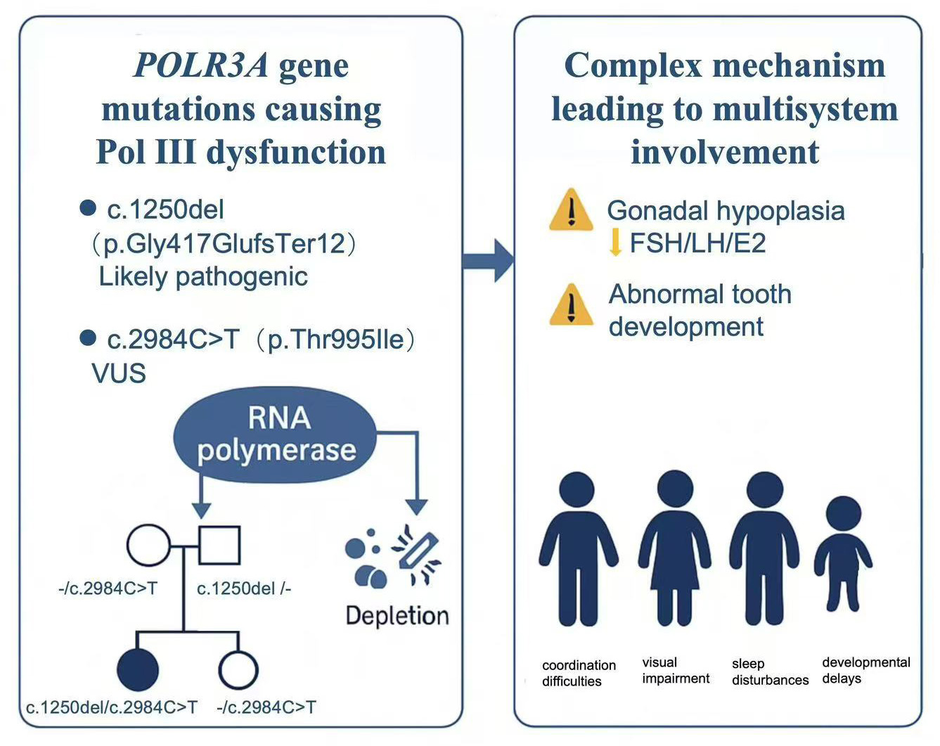
Background:Pathogenic biallelic variants in POLR3A have been associated with different disorders characterized by progressive neurological deterioration. These include the 4H leukodystrophy syndrome (hypomyelination, hypogonadotropic hypogonadism, and hypodontia) and adolescent-onset progressive spastic ataxia, as well as Wiedemann–Rautenstrauch syndrome (WRS), a recognizable neonatal progeroid syndrome. The phenotypic differences between these disorders are thought to occur mainly due to different functional effects of underlying POLR3A variants.
Case presentation: Here, we present the detailed clinical course of a 20-year-old woman who presented with developmental delays, neurological deficits, metabolic abnormalities, and sleep disturbances. She was born to non-consanguineous parents and had an elder sister who usually behaved. Laboratory studies demonstrated low or undetectable LH and FSH levels and abnormally low estradiol levels. MRI showed leukoencephalopathy characterized by white matter lesions and brain atrophy. Homozygous missense mutation c.2984C>T (p.Thr995Ile) was found in POLR3A, which codes for the largest subunit of RNA polymerase Ill.
Conclusions:POLR3A-induced leukodystrophy is relatively rare and not well understood, making it challenging to diagnose and easy to overlook. The prognosis for this disease is generally poor, significantly impacting the quality of life of affected individuals. Since Pol III-related leukodystrophies shows various combination of neurologic and non-neurologic features, additional report will help to bring crucial information concerning this molecular diagnosis, the prediction of the disease and practical consequences for genetic counseling.
Introduction
The POLR3A gene encodes the catalytic subunit of RNA polymerase III (Pol III), a critical enzyme that transcribes a diverse array of Pol III-dependent genes. These genes include structural RNAs, such as 5S rRNA, tRNA, and small nuclear RNAs (e.g., U6 snRNA), as well as other small regulatory RNA molecules, including 7SK snRNA and 7SL RNA[1, 2]. These RNA species play pivotal roles in fundamental cellular processes, including protein synthesis, gene expression regulation, and signal transduction. Mutations in the POLR3A gene lead to RNA polymerase III enzyme dysfunction, ultimately disrupting Pol III-dependent RNA synthesis and metabolism[3]. There are three fundamental pathological mechanisms: (1) Structural RNA deficiency: The reduced Pol III activity impairs the production of essential structural RNAs crucial for protein synthesis[1]; (2) Depletion of small regulatory RNAs: The diminished synthesis of small regulatory RNA molecules disrupts cellular signaling pathways and post-transcriptional regulation[1]; (3) Imbalance in cellular RNA metabolism: The overall dysregulation of RNA homeostasis can lead to cell toxicity and death, indicating fundamental importance of these Pol III-dependent RNAs in cellular viability[3].
Case presentation
A 20-year-old female patient presented with developmental delays (delayed puberty, short stature, and poor academic performance), neurological deficits (visual impairment, hearing preservation, muscle weakness, coordination difficulties, and dental abnormalities), metabolic abnormalities (recurrent infections, skin allergies, and digestive issues), and sleep disturbances (difficulty falling asleep, delayed sleep onset, and insufficient sleep duration), which is consistent with the known function of the POLR3A gene encoding the largest subunit of RNA polymerase III (Pol III). The patient has two heterozygous variations in the POLR3A gene: c.2984C>T (p.Thr995Ile), c.1250del (p.Gly417GlufsTer12). There is currently no relevant literature report on these two variation sites. The results of family verification showed that these two variants originated from their parents (Figure 1a, Figure 1d). Brain Magnetic Resonance Imaging (MRI) confirmed leukoencephalopathy characterized by white matter lesions and brain atrophy (Figure 2). Ultrasound of the patient's gonadal organs showed gonadal hypoplasia (Figure 1c). The Endocrine panel showed endocrine disturbances predominantly involving LH, FSH, and estradiol levels (Table 1). Abnormal tooth development is the main feature of dental abnormality in this patient (Figure 1b).
Figure 1. A Pedigree Chart: Parents are heterozygous, and the daughter has a homozygous mutation. b Schematic diagram of patient's oral cavity and teeth: The patient has abnormal tooth development. c Patient’s ultrasound examinations: Uterine ultrasound (The size of the corpus uteri is 1.5×1.4×1.2cm), Ovarian ultrasound (the size of the left ovary is 2.0×1.0cm, the size of the right ovary is 2.1×1.0cm), Breast ultrasound (The thickness of the left gland is about 0.6cm, the thickness of the right gland is about 0.6cm). d Genetic Sanger sequencing report: patient: there are two heterozygous variations in POLR3A gene: c.2984C>T (p.Thr995Ile), c.1250del (p.Gly417GlufsTer12), patient's father: there is a heterozygous variation at the c.1250del site and no variation is found at the c.2984C>T site, patient's mother: there is a heterozygous variation at the c.2984C>T site and no variation is found at the c.1250del site, patient's elder sister: there is a heterozygous variation at the c.2984C>T site and no variation is found at the c.1250del site.
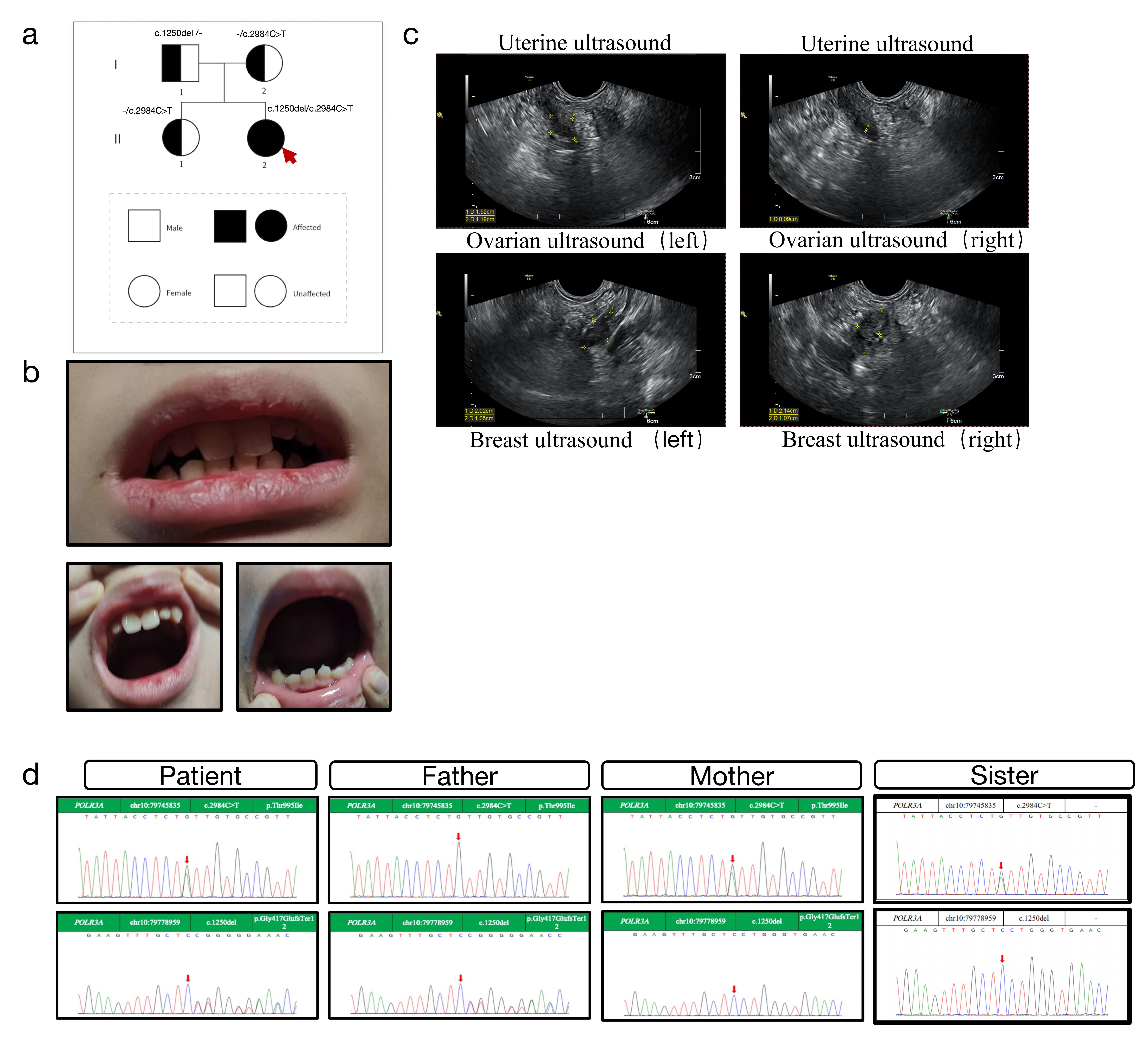
Figure 2. Brain MRI shows brain white matter lesions and brain atrophy, and the pituitary MRI appears generally normal.
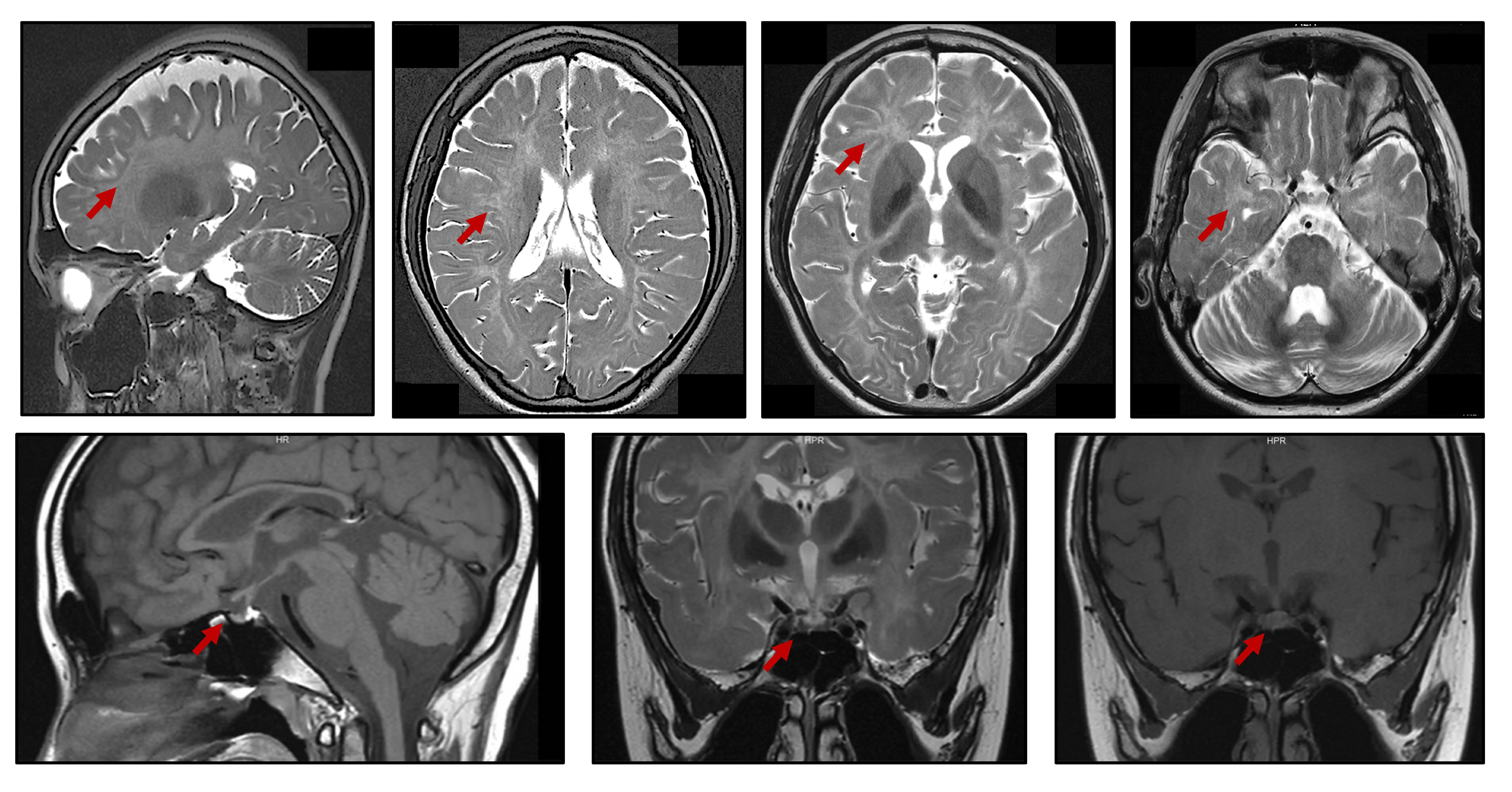
Table 1. Pituitary-Gonad Hormones Profile
|
Item |
Number value |
Property |
|
PRL |
5.38 ng/ml |
normal |
|
GH |
2.28 ng/ml |
normal |
|
COR |
153 ng/ml |
normal |
|
LH |
N.D mlU/ml |
N.D |
|
FSH |
N.D mlU/ml |
N.D |
|
E2 |
11.8 pg/ml |
abnomal |
|
P4 |
N.D ng/ml |
N.D |
|
T |
0.22 ng/ml |
normal |
N.D: Not Detected
The results of the patient's endocrine panel: PRL (Prolactin): 5.38 ng/ml, within the normal range, GH (Growth Hormone): 2.28 ng/ml, within the normal range, COR (Cortisol): 153 ng/ml, within the normal range, LH (Luteinizing Hormone): Not Detected (N.D.) mIU/ml, FSH (Follicle-Stimulating Hormone): Not Detected (N.D.) mIU/ml, E2 (Estradiol): 11.8 pg/ml, abnormal (low), P4 (Progesterone): Not Detected (N.D.) ng/ml, T (Testosterone): 0.22 ng/ml, within the normal range, The low or undetectable levels of LH and FSH, along with the abnormally low estradiol level, suggest a possible dysfunction or deficiency in the patient's reproductive hormones. This could be related to the reproductive organ abnormalities mentioned in the previous information. Further clinical evaluation and interpretation by a medical professional would be necessary to determine these findings’ underlying cause and clinical significance.
Discussion
The patient's comprehensive clinical manifestations, encompassing developmental delays, multisystem neurological deficits, metabolic abnormalities, sleep disturbances, and specific genetic abnormality, are highly suggestive of POLR3A-related leukodystrophy (also known as 4H leukodystrophy syndrome). The hallmark neurological feature of POLR3A-related leukodystrophy is progressive leukoencephalopathy, typically seen in early childhood3. Patients with POLR3A-related leukodystrophy often exhibit delayed motor development, accompanied by ataxia, spasticity, and other movement disorders. Cognitive impairment and developmental delays are also common[3]. Endocrine abnormality is a hallmark of POLR3A-related leukodystrophy. Affected individuals frequently present with delayed or incomplete puberty[1]. Other endocrine disturbances, including growth hormone deficiency and hypothyroidism, have been reported[4]. Dental anomalies, including hypodontia, microdontia, delayed tooth eruption, and enamel hypoplasia, are often present. Additionally, some individuals have been reported to have orofacial dysmorphic features, including a high-arched palate and retrognathia[5].
Neurological manifestations, including visual impairment, hearing impairment, muscle weakness, and coordination difficulties, are typically associated with impaired hypomyelinations caused by POLR3A gene mutations. Susceptibility to infections, skin allergies, digestive issues, and dental problems may be related to the disruption in cellular metabolism caused by POLR3A gene dysfunction. Sleep disturbances may be associated with sleep-wake cycle dysregulation due to central nervous system involvement in POLR3A-related leukodystrophy. Furthermore, Emerging evidence suggests that the specific phenotypic manifestations may be influenced by the nature and location of the underlying POLR3A mutations[6]. Certain mutations have been associated with more severe neurological manifestations, while others may result in milder or atypical clinical course. The diverse phenotypes of POLR3A-related disorders and underlying mechanisms need to be further investigated in the future.
Conclusion
This case underscores the importance of early diagnosis and genetic counseling for POLR3A-related leukodystrophy, characterized by multisystem involvement and neurological deficits. Further investigation is needed to clarify the genotype-phenotype correlations and underlying disease mechanisms.
Acknowledgements
We would like to thank the patient and their families for providing us with detailed information and cooperating with all the medical examinations.
Competing Interests
The authors declare that they have no existing or potential commercial or financial relationships that could create a conflict of interest at the time of conducting this study.
Data Availability
All data needed to evaluate the conclusions in the paper are present in the paper or the Supplementary Materials. Additional data related to this paper may be requested from the authors.
References
[1] Tetreault M, Choquet K, Orcesi S, Tonduti D, Balottin U, Teichmann M, et al. Recessive mutations in POLR3B, encoding the second largest subunit of Pol III, cause a rare hypomyelinating leukodystrophy. Am J Hum Genet. 2011;89(5):652-5. URL: https://www.cell.com/ajhg/fulltext/S0002-9297(11)00440-X
[2] Saitsu H, Osaka H, Sasaki M, Takanashi J, Hamada K, Yamashita A, et al. Mutations in POLR3A and POLR3B encoding RNA Polymerase III subunits cause an autosomal-recessive hypomyelinating leukoencephalopathy. Am J Hum Genet. 2011;89(5):644-51.
[3] McKenna MC, O'Connor A, Lockhart A, Bogdanova-Mihaylova P, Brett F, Langan Y, et al. POLR3A-related disorders: expanding the clinical phenotype. J Neurol. 2024;271(6):3635-8. URL: https://link.springer.com/article/10.1007/s00415-024-12265-9
[4] Wolf NI, Vanderver A, van Spaendonk RM, Schiffmann R, Brais B, Bugiani M, et al. Clinical spectrum of 4H leukodystrophy caused by POLR3A and POLR3B mutations. Neurology. 2014;83(21):1898-905. URL: https://www.neurology.org/doi/10.1212/WNL.0000000000001002?url_ver=Z39.88-2003&rfr_id=ori:rid:crossref.org&rfr_dat=cr_pub%20%200pubmed
[5] Harting I, Al-Saady M, Krageloh-Mann I, Bley A, Hempel M, Bierhals T, et al. POLR3A variants with striatal involvement and extrapyramidal movement disorder. Neurogenetics. 2020;21(2):121-33. URL: https://link.springer.com/article/10.1007/s10048-019-00602-4
[6] Thiffault I, Wolf NI, Forget D, Guerrero K, Tran LT, Choquet K, et al. Recessive mutations in POLR1C cause a leukodystrophy by impairing biogenesis of RNA polymerase III. Nat Commun. 2015;6:7623. URL: https://www.nature.com/articles/ncomms8623
 Figures
 References
 Peer
Peer Information
Figure 1. A Pedigree Chart: Parents are heterozygous, and the daughter has a homozygous mutation. b Schematic diagram of patient's oral cavity and teeth: The patient has abnormal tooth development. c Patient’s ultrasound examinations: Uterine ultrasound (The size of the corpus uteri is 1.5×1.4×1.2cm), Ovarian ultrasound (the size of the left ovary is 2.0×1.0cm, the size of the right ovary is 2.1×1.0cm), Breast ultrasound (The thickness of the left gland is about 0.6cm, the thickness of the right gland is about 0.6cm). d Genetic Sanger sequencing report: patient: there are two heterozygous variations in POLR3A gene: c.2984C>T (p.Thr995Ile), c.1250del (p.Gly417GlufsTer12), patient's father: there is a heterozygous variation at the c.1250del site and no variation is found at the c.2984C>T site, patient's mother: there is a heterozygous variation at the c.2984C>T site and no variation is found at the c.1250del site, patient's elder sister: there is a heterozygous variation at the c.2984C>T site and no variation is found at the c.1250del site.
Figure 2. Brain MRI shows brain white matter lesions and brain atrophy, and the pituitary MRI appears generally normal.
[1] Tetreault M, Choquet K, Orcesi S, Tonduti D, Balottin U, Teichmann M, et al. Recessive mutations in POLR3B, encoding the second largest subunit of Pol III, cause a rare hypomyelinating leukodystrophy. Am J Hum Genet. 2011;89(5):652-5. URL: https://www.cell.com/ajhg/fulltext/S0002-9297(11)00440-X
[2] Saitsu H, Osaka H, Sasaki M, Takanashi J, Hamada K, Yamashita A, et al. Mutations in POLR3A and POLR3B encoding RNA Polymerase III subunits cause an autosomal-recessive hypomyelinating leukoencephalopathy. Am J Hum Genet. 2011;89(5):644-51.
[3] McKenna MC, O'Connor A, Lockhart A, Bogdanova-Mihaylova P, Brett F, Langan Y, et al. POLR3A-related disorders: expanding the clinical phenotype. J Neurol. 2024;271(6):3635-8. URL: https://link.springer.com/article/10.1007/s00415-024-12265-9
[4] Wolf NI, Vanderver A, van Spaendonk RM, Schiffmann R, Brais B, Bugiani M, et al. Clinical spectrum of 4H leukodystrophy caused by POLR3A and POLR3B mutations. Neurology. 2014;83(21):1898-905. URL: https://www.neurology.org/doi/10.1212/WNL.0000000000001002?url_ver=Z39.88-2003&rfr_id=ori:rid:crossref.org&rfr_dat=cr_pub%20%200pubmed
[5] Harting I, Al-Saady M, Krageloh-Mann I, Bley A, Hempel M, Bierhals T, et al. POLR3A variants with striatal involvement and extrapyramidal movement disorder. Neurogenetics. 2020;21(2):121-33. URL: https://link.springer.com/article/10.1007/s10048-019-00602-4
[6] Thiffault I, Wolf NI, Forget D, Guerrero K, Tran LT, Choquet K, et al. Recessive mutations in POLR1C cause a leukodystrophy by impairing biogenesis of RNA polymerase III. Nat Commun. 2015;6:7623. URL: https://www.nature.com/articles/ncomms8623
Peer-review Terminology
Identity transparency: Single anonymized
Reviewer interacts with: Editor
Details
This is an open access article under the terms of the Creative Commons Attribution License(http://creativecommons.org/licenses/by/4.0/), which permits use, distribution and reproduction in any medium, provided the original work is properly cited.
Publication History
Received 2025-01-05
Accepted 2025-03-16
Published 2025-03-30