




Abstract
Background: Chronic rhinosinusitis (CRS) is a complex disease influenced by various factors such as environmental exposure, microbial infection, immune dysfunction, and genetic predisposition. The objective of this research is to examine the causal relationship between plasma lipidomes and CRS, focusing on the potential role of immune cells as mediators.
Methods: Utilizing pooled data from genome-wide association studies (GWAS), a two-sample Mendelian randomization (MR) analysis was conducted to investigate the causal relationship between genetically predicted plasma lipidomes (7174 cases) and CRS (7529 cases, 444966 controls). Various methods such as inverse variance weighted, maximum likelihood, MR-Egger, weighted median, weighted mode, and Wald ratio were employed to assess causality. Multiple sensitivity analyses were also conducted to ensure the reliability of the MR results. Furthermore, mediation analysis was used to identify immune cell-mediated pathways linking plasma lipidomes to chronic sinusitis.
Results:The study identified 30 plasma lipidomes associated with chronic sinusitis, with only one remaining significant after FDR correction, namely Phosphatidylinositol (18:1_18:2). Additionally, 52 types of immune cells were found to be related to the disease, with only 3 types remaining significant after FDR correction: CD27+/IgD- CD38+ B cells, CD3+/CD28+ CD4+ T cells, and HLA DR+/CD33+ HLA DR+ CD14- cells. Mediation analysis revealed two distinct mediating relationships, indicating potential pathways from plasma lipidomes to chronic sinusitis through two specific immune cells. Sensitivity analysis results indicated no heterogeneity or pleiotropy in the study.
Conclusion:The findings of this study reinforce the causal link between plasma lipidomes, immune cells, and chronic sinusitis. Identifying these biomarkers offers fresh insights into the pathogenesis of chronic sinusitis, with implications for improved prevention, diagnosis, and treatment of the condition.
Keywords:Chronic rhinosinusitis, plasma lipidomes, Immune cells, Mendelian randomization, Mediation analysis
Introduction
Chronic rhinosinusitis (CRS) is a complex disease that impacts 5-12% of the overall population. It is marked by persistent inflammation in the nasal passages and paranasal sinuses. CRS is diagnosed when an individual experiences two or more symptoms such as nasal congestion, facial pain, reduced sense of smell for a duration of at least 12 weeks, along with objective evidence of sinus inflammation seen through nasal endoscopy or sinus computed tomography[1] . The occurrence of CRS is associated with various factors, including long-term exposure to air pollutants and smoking[2] . Furthermore, CRS often coexists with chronic conditions such as asthma, allergic rhinitis, and gastroesophageal reflux disease (GERD). These comorbidities may facilitate the onset and progression of CRS through common inflammatory pathways[3] . Numerous studies have identified allergic rhinitis and smoking as potential risk factors for the condition. However, findings have been mixed, with some studies showing only a slight increase in risk. Other studies have explored additional risk factors including genetic variation, gender, race, air pollution, gastroesophageal reflux disease, and autoimmune diseases[3-5] . The variability in results seen in these epidemiological studies could be due in part to confounding variables and the possibility of reverse causation.
Recent evidence suggests an increasing role for plasma lipidomes in managing CRS[6] . Oxidative modification of lipids occurs during inflammation, resulting in the formation and accumulation of bioactive lipid oxidation products. These products induce specific cellular responses, which regulate the inflammatory process and can influence the outcome of the body's response in acute inflammation during host defense[7] . In CRSwNP, local tissues exhibit significant eosinophilic infiltration, accompanied by marked elevations in Th2-type cytokines such as IL-4, IL-5, and IL-13[8] . These cytokines promote the recruitment and activation of inflammatory cells by activating the PI3K/Akt signaling pathway, leading to tissue edema, increased mucus secretion, and impaired epithelial barrier function[9] . Furthermore, the tissue remodeling processes, such as collagen deposition and basement membrane thickening, exacerbate the obstruction of the sinus cavity and perpetuate chronic inflammation[10] . The immune cell-mediated pathway from plasma lipidomes to CRS remains largely unexplored. In recent years, with the development of high-throughput sequencing technology, significant progress has been made in the genetic research of CRS. Several Genome-Wide Association Studies (GWAS) have identified multiple gene loci that are significantly associated with susceptibility to CRS, such as HLCS , HLA-DRA, BICD2, VSIR and SLC5A1[11,12] . In recent years, the interaction between lipid metabolism and inflammatory responses has become a focal point in the study of Cytokine Release Syndrome (CRS). Lipid oxidation products, such as oxidized phospholipids and eicosanoids, not only act as inflammatory mediators involved in the recruitment and activation of immune cells but also influence the intensity and duration of the inflammatory response by regulating signaling pathways such as PI3K/Akt and NF-κB[7] . Furthermore, metabolites of phosphatidylinositol (PI) play a crucial role in modulating immune cell functions, including T cell activation and B cell antibody production[13] . These findings suggest that abnormalities in lipid metabolism may contribute to the onset and progression of CRS by affecting immune cell functions. Mendelian randomization (MR) analysis is a powerful method that leverages genetic variation as an instrumental variable (IV) to investigate potential causal relationships between exposures and outcomes. By using genetic variants that are randomly assigned at conception, MR minimizes the influence of confounding factors on causal estimates. Additionally, mediation analysis can be employed to examine how exposure affects an outcome through mediators. In this study, we conducted MR analysis using publicly available GWAS summary data to explore the causal link between plasma lipidomes, immune cells, and chronic sinusitis, as well as to investigate the immune cell-mediated pathway leading from plasma lipidomes to chronic sinusitis.
Materials and methods
Study design
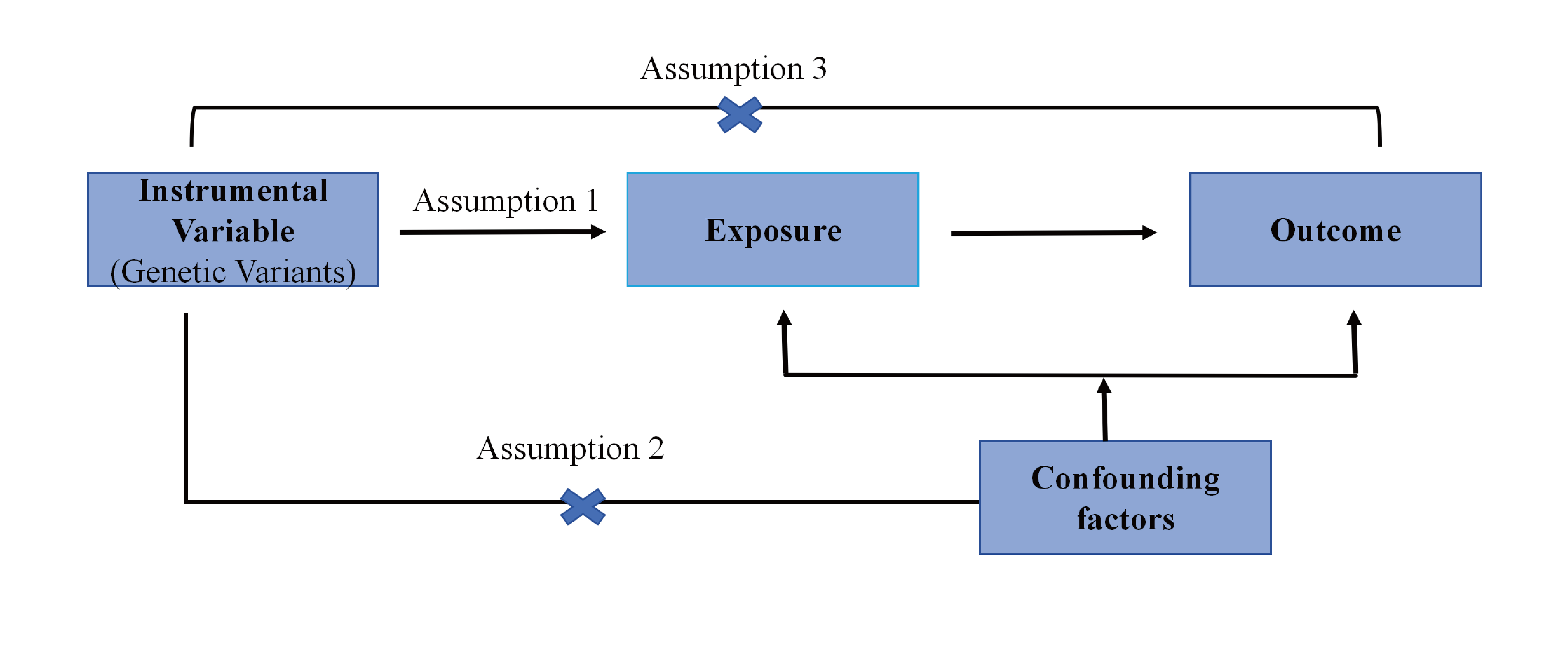
The research flow chart is illustrated in Figure 1. Initially, we acquired published GWAS summary data containing variables such as plasma lipidomes, immune cells, and CRS. Subsequently, two-sample MR analysis was employed to assess the causal association between plasma lipidomes, immune cells, and CRS. Lastly, a two-step assay was conducted to ascertain the mediating effect of immune cells in the connection between plasma lipidomes and CRS. Our MR investigations adhere to the STROBE-MR guidelines[14] .
Figure 1. Mendelian randomization mediation analysis flowchart
Data sources
Summary statistics for plasma lipidomes were obtained from the GWAS catalog (https://www.ebi.ac.uk/gwas/) under study accession numbers GCST90277238-GCST90277416. The study included 13 out of 7174 Finnish individuals from the GeneRISK cohort and conducted univariate and multivariate GWAS on 179 lipid species across different lipid classes.
The public catalog of immune cell GWAS data sources (GCST0001391 to GCST0002121) includes GWAS data for 731 immune phenotypes analyzed in the study.[15] A total of 3,757 Sardinian individuals were analyzed to identify around 22 million genetic variants and investigate their associations with autoimmune diseases using GWAS immunological signature data. The dataset comprised 118 absolute cell counts , 389 median fluorescence intensity (MFI) values reflecting surface antigen levels, 32 morphological features , and 192 relative cell counts , which represent ratios at the cellular level. The GWAS dataset for CRS (identifier: ebi-a-GCST90018823) included 7,529 cases and 444,966 controls.
Genetic IV selection
In order to utilize genetic variation for estimating causal effects, three fundamental assumptions of IV must be satisfied: 1) IV must be associated with exposure factors; 2) IV should not be correlated with confounding factors; 3) IV should not have a direct impact on outcome variables, influencing them solely through exposure factors. The IVs employed in this research were carefully selected based on the following criteria: 1) single nucleotide polymorphisms (SNPs) meeting a site-wide threshold of P < 5×10⁻⁵ were considered when there were limited genome-wide significant sites in the original GWAS results, with a genome-wide significance threshold of P < 5×10⁻⁸ used as potential instrumental variables linked to each exposure trait. 2) SNPs correlated with outcome variables (P < 0.05) were excluded. 3) An aggregation process was conducted to mitigate the effects of linkage disequilibrium (r2 < 0.001, window size = 10,000 kb). 4) The The Mendelian randomization pleiotropy residual sum and outlier (MR-PRESSO) method was employed to assess and address potential pleiotropy, with outliers removed to mitigate pleiotropy effects[16] . SNPs were arranged in ascending order based on the P value of the MR-PRESSO outlier test, with subsequent removal of SNPs until no pleiotropy was detected (MRPRESSO global test P value > 0.05). To assess the strength of the selected SNPs, an F statistic was utilized, excluding SNPs with an F statistic < 10 to mitigate weak instrument bias in MR analysis. The F statistic is calculated using the formula F = [R2 × (n - k - 1)] / [k × (1 - R2)] , where R2 represents the proportion of exposure variance explained by the IV, n is the sample size, and k is the number of IVs[17] .
Mendelian randomization and statistical analysis
Two-sample Mendelian randomization employed MR methods to assess the causal association between plasma lipidomes, immune cells, and CRS. The Wald ratio was utilized to determine causality of exposures, incorporating only a single IV. In cases of exposures with multiple IVs, methods such as inverse variance weighting (IVW), Simple mode, MR-Egger, weighted median, and weighted mode were employed to infer causal relationships. IVW typically offers the highest statistical power and is therefore preferred, with other methods being used as supplementary. IVW utilizes meta-analysis to combine Wald ratio estimates for each IV while constraining the intercept to zero[18] . In the absence of horizontal pleiotropy, the IVW method can yield unbiased causal estimates[19] . In cases of heterogeneity, the random-effects IVW test is preferred for its conservative and robust estimate, while the fixed-effects model is utilized when heterogeneity is absent. MR-Egger is able to detect multiple-level effects and provide valid causal estimates even in the presence of pleiotropy[20] . The weighted median method can yield reliable causal estimates even when as many as 50% of instrumental variables are invalid[21] . If most IVs with similar causal estimates are valid instruments, the weighted pattern approach remains valid even if other IVs do not meet the requirements for MR analysis. It is recommended to conduct a sensitivity analysis to evaluate the robustness of the causal relationship[22] . MR-Egger regression and MR-PRESSO were utilized to evaluate horizontal pleiotropy. The presence of a nonzero intercept in the MR-Egger regression suggests directional pleiotropy. Heterogeneity among instrumental variables (IVs) was assessed using Cochran's Q test. Furthermore, leave-one-out sensitivity analysis was conducted to determine if a single SNP influenced the causal estimate. A false discovery rate (FDR) correction was applied to the primary IVW results using the Benjamini-Hochberg procedure. FDR significance threshold of DR < 0.05 indicates a significant association. All Mendelian randomization analyses were conducted using R software (version 4.3.1) with the 'TwoSampleMR' package (https://github.com/MRCIEU/TwoSampleMR) and the 'MR-PRESSO' package (https://github.com/rondolab/MR-PRESSO).
Reverse Mendelian Randomization
To investigate the potential causal effect of CRS on the identified plasma lipidomes (PIVW < 0.05), a reverse MR analysis was conducted. In this analysis, SNPs associated with CRS were treated as IVs, CRS was considered the exposure, and plasma lipidomes that tested positive in the forward analysis were viewed as outcomes. The methodology for reverse MR analysis closely mirrors that of traditional MR analysis.
Mediation analysis
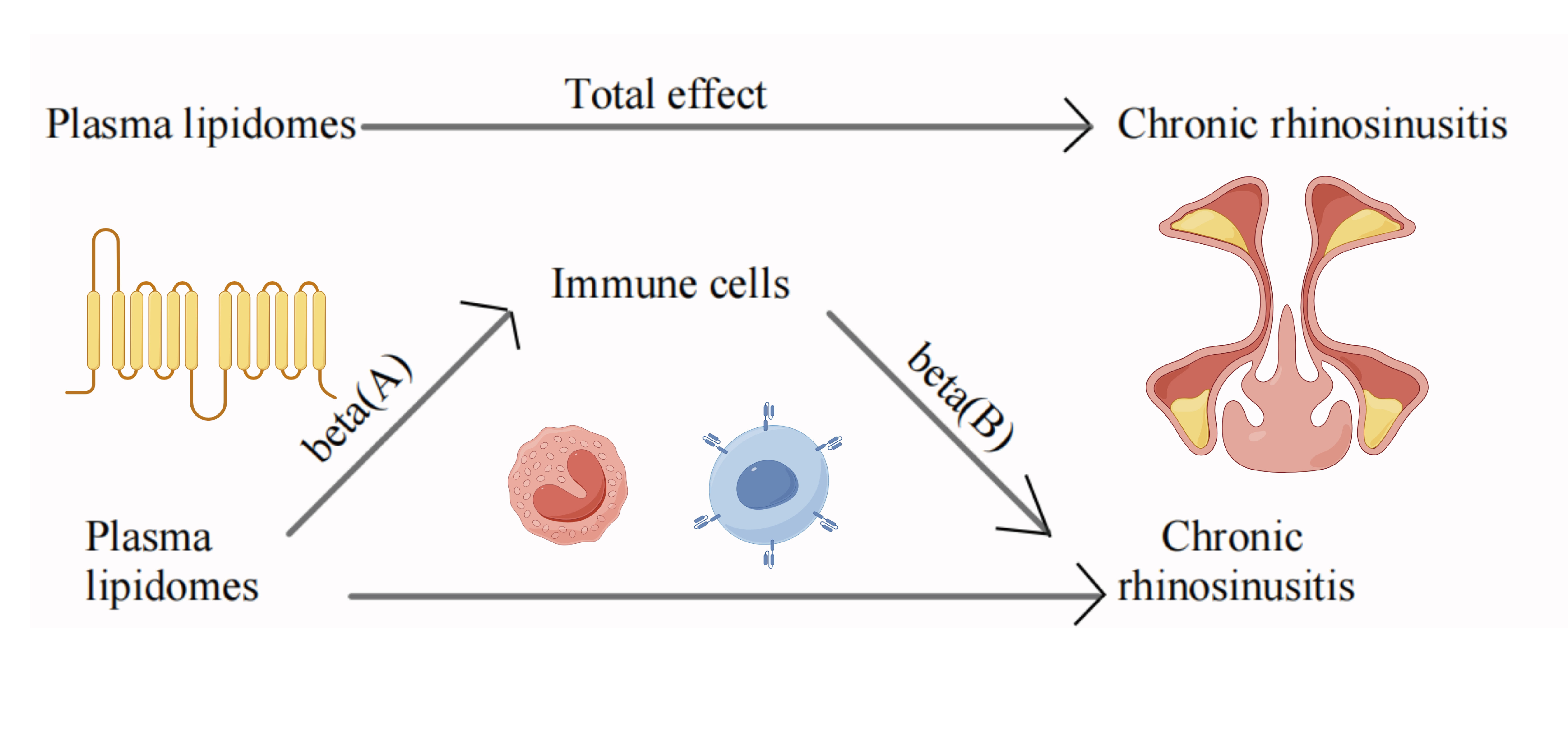
Mediation analysis aims to evaluate the pathway from exposure to outcome through mediators, providing insight into the underlying mechanisms through which exposure influences outcomes[23] . The mediation analysis in this study focused on the relationship between plasma lipidomes and immune cells in the context of CRS. Initially, a two-sample MR approach was employed to assess the causal connection between plasma lipidomes and immune cells, resulting in β(A). Subsequently, a two-step process was utilized to identify immune cells that remained causally linked to CRS even after adjusting for plasma lipidomes, represented by β(B), and to ensure that the mediating effects on outcomes were independent of exposure. The mediation effect was then calculated using a two-step MR method: Mediation effect = β(A) × β(B). The total effect of plasma lipidomes on CRS was determined through the previous two-sample MR analysis, with the direct effect being calculated as (total effect - mediation effect). Finally, the mediation ratio was calculated using the formula: Mediation ratio = (mediation effect/total effect) × 100%.The Bootstrap mediation effect test was utilized to assess the mediation effect[24] .
Results
Causal effects of plasma lipidomes on CRS
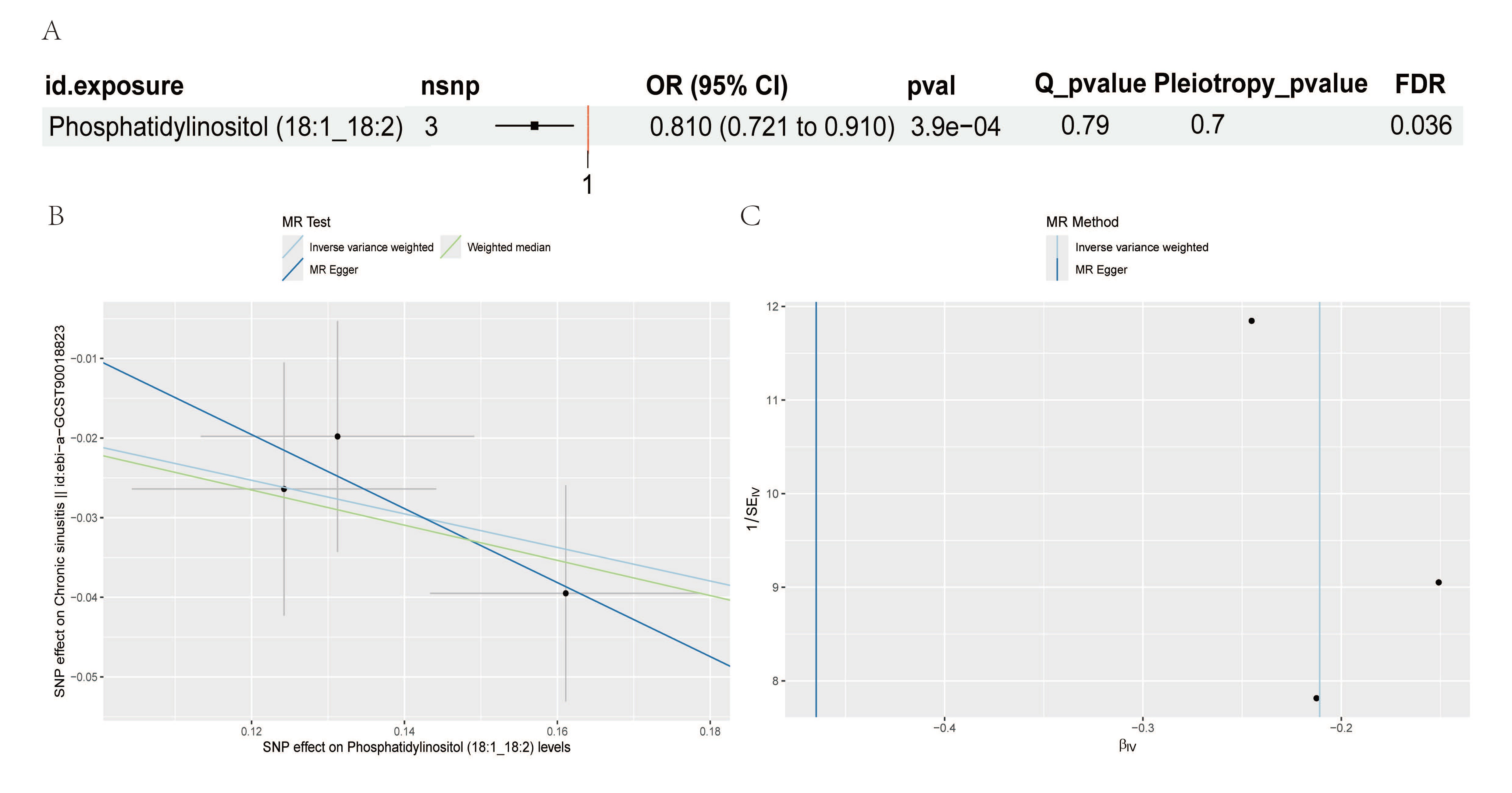
Two sample Mendelian randomization analyses revealed 30 suggestive associations between plasma lipidomes and CRS (Supplementary Table 1). Following correction for false discovery rate (FDR), only Phosphatidylinositol (18:1_18:2) remained significant. Our analysis indicated a significant association between elevated levels of Phosphatidylinositol (18:1_18:2) and a reduced risk of CRS (OR=0.81, 95%CI=0.72-0.91, p=3.9e−04). Cochran's Q test showed no heterogeneity (p = 0.79). Additionally, the pleiotropy test using Egger's intercept demonstrated that our Mendelian randomization study did not exhibit pleiotropy (p = 0.7) ( Figure 2 ).
Figure 2. MR analysis results of the causal relationship between plasma lipidomes on the risk of chronic rhinosinusitis (A) The results of the forest map (B) The result of the scatter plot (C) The result of the funnel plot
Causal effects of immune cells on CRS
The IVW method identified 53 associations between immune cells and CRS (Supplementary Table 2). Following FDR correction, only 3 associations remained significant: CD27 on IgD- CD38+ B cell (OR=1.140, 95%CI=1.060 to 1.226, p=4.1 e−04), CD3 on CD28+ CD4+ T cell (OR=0.93, 95%CI=0.90 to 0.97, p=3e−04), and HLA DR on CD33+ HLA DR+ CD14- (OR=1.05, 95%CI=1.02 to 1.07, p=7e−04). Sensitivity analysis indicated no heterogeneity or horizontal pleiotropy in the CD27 on IgD- CD38+ B cell and CD3 on CD28+ CD4+ T cell analyses. However, the CD3 on CD28+ CD4+ T cell Cochran's Q test revealed significant heterogeneity (p = 4.03e-05), which did not impact the IVW method results. The conclusions drawn are deemed reliable. Additionally, MR Egger's pleiotropy test showed no evidence of pleiotropy in our findings (p = 0.54) (Figure 3).
Figure 3. Forest plot of MR analyses for the causal effects of immune cells on the risk of chronic rhinosinusitis

Reverse two-sample MR analysis
In the Two-Sample Mendelian Randomization (TSMR) analysis comparing Phosphatidylinositol (18:1_18:2) and CRS, a relaxed threshold of 1 × 10⁻⁴ was utilized to increase the number of IVs. However, an insufficient number of exposure SNPs were obtained. A Steiger test was conducted, yielding results indicating correct_causal_direction=TRUE and steiger_pval=9×10⁻³. This suggests that the direction test of individual SNPs is accurate, the overall SNP test direction is correct, and the instrumental variables show no evidence of reverse causality (Supplementary Table 3)[25] .
Mediation analysis results
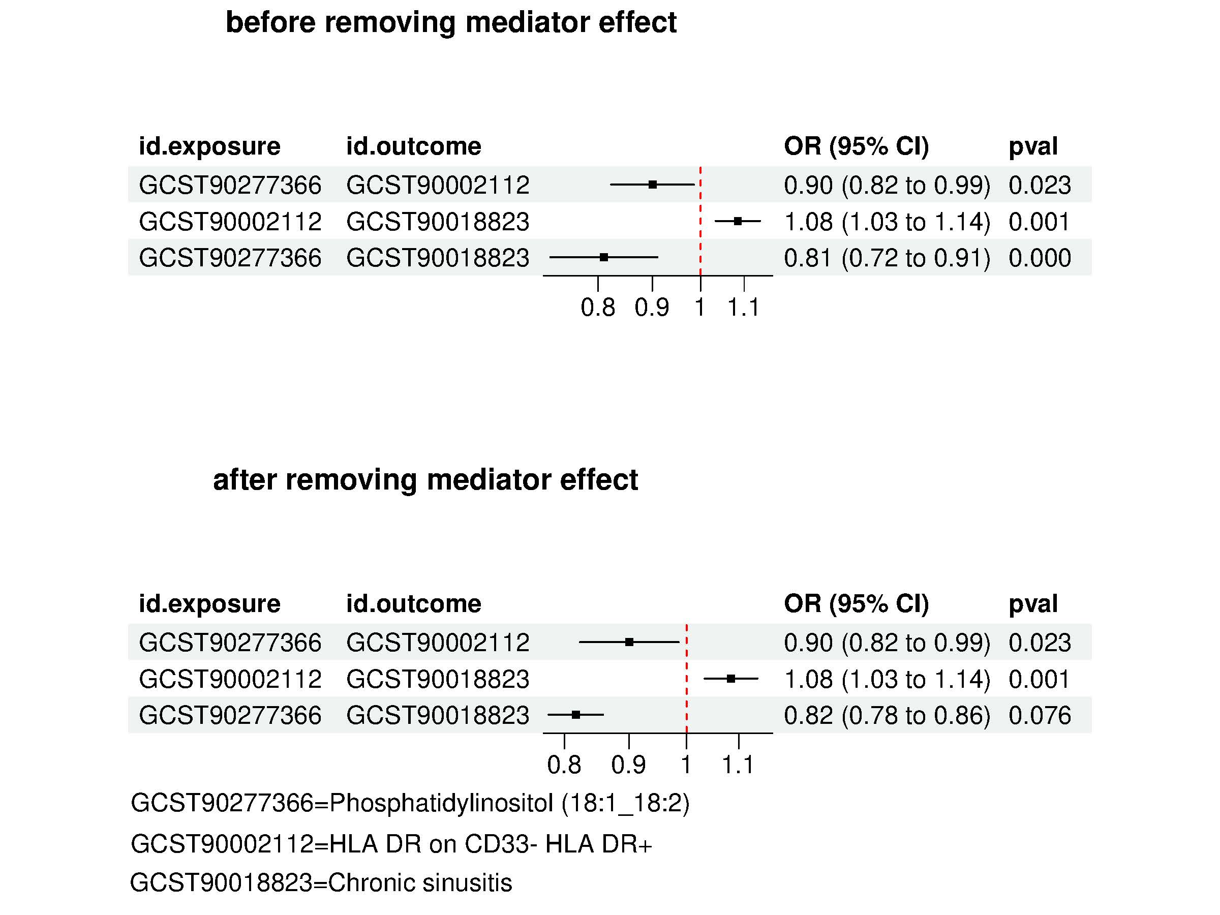
To investigate the mechanisms underlying CRS occurrence and development, we conducted a two-step mediation analysis to uncover the causal pathway between immune cell-mediated Phosphatidylinositol (18:1_18:2) and CRS. This analysis, detailed in the Mediation Analysis section of Methods, focused on CRS-associated plasma lipidomes previously identified in two MR immune cell samples. Initially, the dual-sample MR assessed the causal relationship between these plasma lipidomes and immune cells, revealing 29 associations of Phosphatidylinositol (18:1_18:2) with immune cells(Supplementary Table 4). Subsequently, we examined 29 immune cells as mediators of the liposome-to-CRS pathway. Our findings identified two mediators: Basophil %CD33dim HLA DR- CD66b- (β= -0.002879126; proportional mediation: 1.3%)(Figure 4) and HLA DR on CD33- HLA DR+ (β=-0.008528472; Proportionally mediated: 4%)(Figure 5). These results indicate that the total effect, indirect effect, and direct effect align in the same direction, with leave-one-out analysis supporting the reliability of the conclusion. By recalculating the p value using the bootstrap algorithm, we found that while the original p value from exposure to outcome remains significant, removing the mediating effect renders it insignificant, suggesting that the outcome is indirectly influenced by the mediating effect.(Supplementary Table 5).
Figure 4. Mediation effect of Phosphatidylinositol (18:1_18:2) on chronic rhinosinusitis via Basophil %CD33dim HLA DR- CD66b-
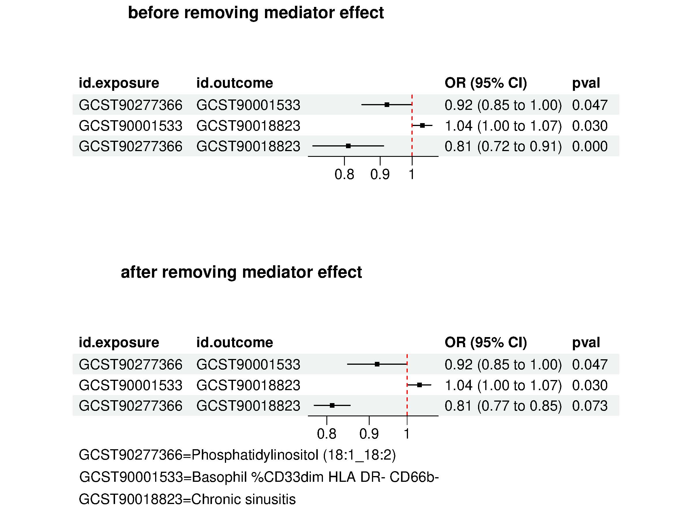
Figure 5 Mediation effect of Phosphatidylinositol (18:1_18:2) on chronic rhinosinusitis via HLA DR on CD33- HLA DR+
Discussion
This study utilized Mendelian randomization analysis to comprehensively assess the causal relationship between plasma lipidomes, immune cells, and CRS. The results indicated potential causal associations between 30 different types of lipids (with only 1 type remaining significant after FDR correction), 52 types of immune system cells (with only 3 types remaining significant after FDR correction), and CRS. Furthermore, genetically predicted Phosphatidylinositol (18:1_18:2) was linked to a reduced risk of CRS, with 1.3% of the effect mediated through Basophil %CD33dim HLA DR- CD66b- and 4% through HLA DR on CD33- HLA DR+. This study is the first to investigate the causal relationship between plasma lipidomes and CRS risk using Mendelian randomization methods, and it also highlights Basophil %CD33dim HLA DR- CD66b- and HLA DR on CD33- HLA DR+ as potential mediators. Robinson et al. point out that lipid mediators play a crucial role in the inflammatory regulation of CRS[6] . Bachert et al. found elevated levels of IL-5 and IgE antibodies in nasal polyp tissues, suggesting the significance of Th2-type immune responses in CRS[10] . Motomura et al. discovered that basophils regulate pulmonary natural helper cells by secreting IL-4, thereby promoting airway inflammation[26] . HLA-DR plays a crucial role in antigen presentation and immune activation, with its expression levels significantly elevated in chronic airway inflammation and correlated with disease severity[27] .
Phosphatidylinositol (18:1_18:2) is a phospholipid, while PI3K is a type of kinase enzyme that acts as a phospholipase. The connection between them lies in the fact that PI3K has the ability to phosphorylate phosphatidylinositol, which in turn plays a role in the regulation of important biological processes like cell growth, proliferation, and survival.
Phosphoinositide 3-kinase-delta may play a role in the development of nasal polyps, particularly eosinophilic nasal polyps which are linked to more severe clinical symptoms and radiographic characteristics[9] . Our study provides evidence of a negative causal relationship between Phosphatidylinositol (18:1_18:2) and CRS.
Phosphoinositide 3-kinase-delta is crucial in modulating adaptive immune responses, particularly in T cells and B cells[28,29] . Phosphatidylinositol (PI) metabolites have the potential to influence the inflammatory response. These metabolites can act as signaling molecules within cells, impacting the function of immune cells and the production of inflammatory mediators[13] . The relationship between immune cells and CRS is intricate. Our research indicates that Basophil %CD33dim HLA DR- CD66b-, HLA DR on CD33- HLA DR+ plays a crucial role as a mediator in the interaction between plasma lipidomes and CRS. Previous studies have extensively explored the connection between basophils and airway inflammation. Basophils are a component of the immune system, contributing to allergic reactions and defense against parasites. They are crucial in the inflammatory response triggered by parasites and allergens[30] . Chronic inflammatory diseases may involve specific immune cell subsets that play a significant role, such as the role of CD66b+ monocytes in inflammation[31] . Basophil-associated OX40 ligand is crucial for the initiation of Th2 responses in airway inflammation,Basophil IL-4 plays a crucial role in the generation of NH-derived cytokines and chemokines, ultimately leading to proteolytic allergen-induced airway inflammation[26] . Basophil IL-4 plays a crucial role in the generation of NH-derived cytokines and chemokines, ultimately leading to proteolytic allergen-induced airway inflammation. HLA-DR, also known as human lymphocyte antigen D-related antigen, is linked to immune irregularities and plays a crucial role in various autoimmune and neurological disorders[27] . HLA-DR gene variants are linked to a higher risk of specific neuropsychiatric disorders. Individuals with conditions like major depressive disorder, sleep disorders, and autism spectrum disorders tend to exhibit elevated levels of HLA-DRB1 in the HLA-DR region[27] . The findings indicate that immunologic issues related to CRS may involve basophil percentages and HLA DR-mediated cell subsets. This study has several limitations. First, even if we take steps to identify and eliminate outlier variables, we cannot rule out the possibility that horizontal pleiotropy affects our results. Second, The study used specific GWAS cohorts (Sardinian, Finnish, and Finnish/UK), which may lack genetic heterogeneity and limit the generalizability of the results. Meanwhile, the lack of cross racial validation and population stratification correction may affect the robustness and extrapolation of the results. In the future, the causal relationship between Phosphatidylinositol and immune cell function will be validated in CRS patient tissues or animal models. In addition, by combining multiple omics data, the lipid immune CRS network will be systematically analyzed
Conclusion
Our study identified 2 mediating relationships, specifically Basophil %CD33dim HLA DR- CD66b- and HLA DR on CD33- HLA DR+, which mediate the causal pathway between Phosphatidylinositol (18:1_18:2) and CRS. The findings from our Mendelian randomization analysis provide support for a causal effect of plasma lipidomes on CRS through immune cells. Additionally, our mediation analysis demonstrated that immune cells play a role in mediating the pathway from plasma lipidomes to CRS. These identified plasma lipidomes and immune cells could serve as potential biomarkers for the diagnosis and treatment of CRS, as well as contribute to further understanding the underlying mechanisms of CRS.
Abbreviations
CRS=Chronic rhinosinusitis, MR=Mendelian randomization, IV=instrumental variable, SNPs=single nucleotide polymorphisms, IVW=inverse variance weighting, TSMR=Two-Sample Mendelian Randomization
Declarations
Author contributions
QG - conception and study design, manuscript writing; DD & YLZ - review and edit manuscript; XJQ & SMH - Data collection, analysis and wrtie-up.
Acknowledgements
We would like to thank all the authors who made outstanding contributions to this study.
Funding information
This research was partially funded by the National Natural Science Foundation of China (82071023),the Key Project of Medical Science and Technology of Henan Province (SBGJ202102160), and the Scientific Research Project of Col-leges and Universities in Henan Province (21A320053).
Ethics approval and consent to participate
An ethics statement may not be necessary for this MR study, as it utilizes summary statistics from previously published GWAS studies. The use of publicly available data sources, in this case, does not require the approval of an ethics committee or institutional review board.
Competing Interests
The authors declare that they have no existing or potential commercial or financial relationships that could create a conflict of interest at the time of conducting this study.
Data Availability
All data needed to evaluate the conclusions in the paper are present in the paper or the Supplementary Materials. Additional data related to this paper may be requested from the authors.
References
[1] Fokkens, W.J., et al., European Position Paper on Rhinosinusitis and Nasal Polyps 2020. Rhinology, 2020. 58(Suppl S29): p. 1-464.
[2] Christensen, D.N., et al., A Systematic Review of the Association between Cigarette Smoke Exposure and Chronic Rhinosinusitis. Otolaryngol Head Neck Surg, 2018. 158(5): p. 801-816.
[3] Sedaghat, A.R., E.C. Kuan, and G.K. Scadding, Epidemiology of Chronic Rhinosinusitis: Prevalence and Risk Factors. J Allergy Clin Immunol Pract, 2022. 10(6): p. 1395-1403.
[4] Orlandi, R.R., et al., International Consensus Statement on Allergy and Rhinology: Rhinosinusitis. Int Forum Allergy Rhinol, 2016. 6 Suppl 1: p. S22-209.
[5] Min, J.Y. and B.K. Tan, Risk factors for chronic rhinosinusitis. Curr Opin Allergy Clin Immunol, 2015. 15(1): p. 1-13.
[6] Robinson, P.Z., D.N. Frank, and V.R. Ramakrishnan, Inflammation resolution and specialized pro-resolving lipid mediators in chronic rhinosinusitis. Expert Rev Clin Immunol, 2023. 19(8): p. 969-979.
[7] Bochkov, V.N. and N. Leitinger, Anti-inflammatory properties of lipid oxidation products. J Mol Med (Berl), 2003. 81(10): p. 613-26.
[8] Simmalee, K., et al., Inflammatory and homeostatic roles of eosinophil subpopulations in chronic rhinosinusitis with nasal polyp pathogenesis. Front Immunol, 2025. 16: p. 1568541.
[9] Kim, J.S., et al., Phosphoinositide 3-kinase-delta could be a biomarker for eosinophilic nasal polyps. Sci Rep, 2018. 8(1): p. 15990.
[10] Bachert, C., et al., Presence of IL-5 protein and IgE antibodies to staphylococcal enterotoxins in nasal polyps is associated with comorbid asthma. J Allergy Clin Immunol, 2010. 126(5): p. 962-8, 968.e1-6.
[11] Bohman, A., et al., A family-based genome-wide association study of chronic rhinosinusitis with nasal polyps implicates several genes in the disease pathogenesis. PLoS One, 2017. 12(12): p. e0185244.
[12] Soliai, M., et al., Two-stage genome-wide association study of chronic rhinosinusitis and disease subphenotypes highlights mucosal immunity contributing to risk. Int Forum Allergy Rhinol, 2021. 11(4): p. 814-817.
[13] Nakamura, Y., et al., Phosphatidylinositol-specific phospholipase C enhances epidermal penetration by Staphylococcus aureus. Sci Rep, 2020. 10(1): p. 17845.
[14] Skrivankova, V.W., et al., Strengthening the Reporting of Observational Studies in Epidemiology Using Mendelian Randomization: The STROBE-MR Statement. Jama, 2021. 326(16): p. 1614-1621.
[15] Orrù, V., et al., Author Correction: Complex genetic signatures in immune cells underlie autoimmunity and inform therapy. Nat Genet, 2020. 52(11): p. 1266.
[16] Verbanck, M., et al., Detection of widespread horizontal pleiotropy in causal relationships inferred from Mendelian randomization between complex traits and diseases. Nat Genet, 2018. 50(5): p. 693-698.
[17] Pierce, B.L., H. Ahsan, and T.J. Vanderweele, Power and instrument strength requirements for Mendelian randomization studies using multiple genetic variants. Int J Epidemiol, 2011. 40(3): p. 740-52.
[18] Burgess, S., A. Butterworth, and S.G. Thompson, Mendelian randomization analysis with multiple genetic variants using summarized data. Genet Epidemiol, 2013. 37(7): p. 658-65.
[19] Hemani, G., et al., The MR-Base platform supports systematic causal inference across the human phenome. Elife, 2018. 7.
[20] Bowden, J., G. Davey Smith, and S. Burgess, Mendelian randomization with invalid instruments: effect estimation and bias detection through Egger regression. Int J Epidemiol, 2015. 44(2): p. 512-25.
[21] Bowden, J., et al., Consistent Estimation in Mendelian Randomization with Some Invalid Instruments Using a Weighted Median Estimator. Genet Epidemiol, 2016. 40(4): p. 304-14.
[22] Hartwig, F.P., G. Davey Smith, and J. Bowden, Robust inference in summary data Mendelian randomization via the zero modal pleiotropy assumption. Int J Epidemiol, 2017. 46(6): p. 1985-1998.
[23] Carter, A.R., et al., Mendelian randomisation for mediation analysis: current methods and challenges for implementation. Eur J Epidemiol, 2021. 36(5): p. 465-478.
[24] Rogne, T., et al., Mediating Factors in the Association of Maternal Educational Level With Pregnancy Outcomes: A Mendelian Randomization Study. JAMA Netw Open, 2024. 7(1): p. e2351166.
[25] Hemani, G., K. Tilling, and G. Davey Smith, Correction: Orienting the causal relationship between imprecisely measured traits using GWAS summary data. PLoS Genet, 2017. 13(12): p. e1007149.
[26] Motomura, Y., et al., Basophil-derived interleukin-4 controls the function of natural helper cells, a member of ILC2s, in lung inflammation. Immunity, 2014. 40(5): p. 758-71.
[27] Rohn, H., et al., Effect of HLA-G5 Immune Checkpoint Molecule on the Expression of ILT-2, CD27, and CD38 in Splenic B cells. J Immunol Res, 2022. 2022: p. 4829227.
[28] Okkenhaug, K., et al., Impaired B and T cell antigen receptor signaling in p110delta PI 3-kinase mutant mice. Science, 2002. 297(5583): p. 1031-4.
[29] Okkenhaug, K., et al., The p110delta isoform of phosphoinositide 3-kinase controls clonal expansion and differentiation of Th cells. J Immunol, 2006. 177(8): p. 5122-8.
[30] Vivanco Gonzalez, N., et al., Mass Cytometry Phenotyping of Human Granulocytes Reveals Novel Basophil Functional Heterogeneity. iScience, 2020. 23(11): p. 101724.
[31] Horzum, U., et al., CD66b(+) monocytes represent a proinflammatory myeloid subpopulation in cancer. Cancer Immunol Immunother, 2021. 70(1): p. 75-87.
 Figures
 References
 Peer
Peer Information
Figure 1. Mendelian randomization mediation analysis flowchart
Figure 2. MR analysis results of the causal relationship between plasma lipidomes on the risk of chronic rhinosinusitis (A) The results of the forest map (B) The result of the scatter plot (C) The result of the funnel plot
Figure 3. Forest plot of MR analyses for the causal effects of immune cells on the risk of chronic rhinosinusitis
Figure 4. Mediation effect of Phosphatidylinositol (18:1_18:2) on chronic rhinosinusitis via Basophil %CD33dim HLA DR- CD66b-
Figure 5 Mediation effect of Phosphatidylinositol (18:1_18:2) on chronic rhinosinusitis via HLA DR on CD33- HLA DR+
[1] Fokkens, W.J., et al., European Position Paper on Rhinosinusitis and Nasal Polyps 2020. Rhinology, 2020. 58(Suppl S29): p. 1-464.
[2] Christensen, D.N., et al., A Systematic Review of the Association between Cigarette Smoke Exposure and Chronic Rhinosinusitis. Otolaryngol Head Neck Surg, 2018. 158(5): p. 801-816.
[3] Sedaghat, A.R., E.C. Kuan, and G.K. Scadding, Epidemiology of Chronic Rhinosinusitis: Prevalence and Risk Factors. J Allergy Clin Immunol Pract, 2022. 10(6): p. 1395-1403.
[4] Orlandi, R.R., et al., International Consensus Statement on Allergy and Rhinology: Rhinosinusitis. Int Forum Allergy Rhinol, 2016. 6 Suppl 1: p. S22-209.
[5] Min, J.Y. and B.K. Tan, Risk factors for chronic rhinosinusitis. Curr Opin Allergy Clin Immunol, 2015. 15(1): p. 1-13.
[6] Robinson, P.Z., D.N. Frank, and V.R. Ramakrishnan, Inflammation resolution and specialized pro-resolving lipid mediators in chronic rhinosinusitis. Expert Rev Clin Immunol, 2023. 19(8): p. 969-979.
[7] Bochkov, V.N. and N. Leitinger, Anti-inflammatory properties of lipid oxidation products. J Mol Med (Berl), 2003. 81(10): p. 613-26.
[8] Simmalee, K., et al., Inflammatory and homeostatic roles of eosinophil subpopulations in chronic rhinosinusitis with nasal polyp pathogenesis. Front Immunol, 2025. 16: p. 1568541.
[9] Kim, J.S., et al., Phosphoinositide 3-kinase-delta could be a biomarker for eosinophilic nasal polyps. Sci Rep, 2018. 8(1): p. 15990.
[10] Bachert, C., et al., Presence of IL-5 protein and IgE antibodies to staphylococcal enterotoxins in nasal polyps is associated with comorbid asthma. J Allergy Clin Immunol, 2010. 126(5): p. 962-8, 968.e1-6.
[11] Bohman, A., et al., A family-based genome-wide association study of chronic rhinosinusitis with nasal polyps implicates several genes in the disease pathogenesis. PLoS One, 2017. 12(12): p. e0185244.
[12] Soliai, M., et al., Two-stage genome-wide association study of chronic rhinosinusitis and disease subphenotypes highlights mucosal immunity contributing to risk. Int Forum Allergy Rhinol, 2021. 11(4): p. 814-817.
[13] Nakamura, Y., et al., Phosphatidylinositol-specific phospholipase C enhances epidermal penetration by Staphylococcus aureus. Sci Rep, 2020. 10(1): p. 17845.
[14] Skrivankova, V.W., et al., Strengthening the Reporting of Observational Studies in Epidemiology Using Mendelian Randomization: The STROBE-MR Statement. Jama, 2021. 326(16): p. 1614-1621.
[15] Orrù, V., et al., Author Correction: Complex genetic signatures in immune cells underlie autoimmunity and inform therapy. Nat Genet, 2020. 52(11): p. 1266.
[16] Verbanck, M., et al., Detection of widespread horizontal pleiotropy in causal relationships inferred from Mendelian randomization between complex traits and diseases. Nat Genet, 2018. 50(5): p. 693-698.
[17] Pierce, B.L., H. Ahsan, and T.J. Vanderweele, Power and instrument strength requirements for Mendelian randomization studies using multiple genetic variants. Int J Epidemiol, 2011. 40(3): p. 740-52.
[18] Burgess, S., A. Butterworth, and S.G. Thompson, Mendelian randomization analysis with multiple genetic variants using summarized data. Genet Epidemiol, 2013. 37(7): p. 658-65.
[19] Hemani, G., et al., The MR-Base platform supports systematic causal inference across the human phenome. Elife, 2018. 7.
[20] Bowden, J., G. Davey Smith, and S. Burgess, Mendelian randomization with invalid instruments: effect estimation and bias detection through Egger regression. Int J Epidemiol, 2015. 44(2): p. 512-25.
[21] Bowden, J., et al., Consistent Estimation in Mendelian Randomization with Some Invalid Instruments Using a Weighted Median Estimator. Genet Epidemiol, 2016. 40(4): p. 304-14.
[22] Hartwig, F.P., G. Davey Smith, and J. Bowden, Robust inference in summary data Mendelian randomization via the zero modal pleiotropy assumption. Int J Epidemiol, 2017. 46(6): p. 1985-1998.
[23] Carter, A.R., et al., Mendelian randomisation for mediation analysis: current methods and challenges for implementation. Eur J Epidemiol, 2021. 36(5): p. 465-478.
[24] Rogne, T., et al., Mediating Factors in the Association of Maternal Educational Level With Pregnancy Outcomes: A Mendelian Randomization Study. JAMA Netw Open, 2024. 7(1): p. e2351166.
[25] Hemani, G., K. Tilling, and G. Davey Smith, Correction: Orienting the causal relationship between imprecisely measured traits using GWAS summary data. PLoS Genet, 2017. 13(12): p. e1007149.
[26] Motomura, Y., et al., Basophil-derived interleukin-4 controls the function of natural helper cells, a member of ILC2s, in lung inflammation. Immunity, 2014. 40(5): p. 758-71.
[27] Rohn, H., et al., Effect of HLA-G5 Immune Checkpoint Molecule on the Expression of ILT-2, CD27, and CD38 in Splenic B cells. J Immunol Res, 2022. 2022: p. 4829227.
[28] Okkenhaug, K., et al., Impaired B and T cell antigen receptor signaling in p110delta PI 3-kinase mutant mice. Science, 2002. 297(5583): p. 1031-4.
[29] Okkenhaug, K., et al., The p110delta isoform of phosphoinositide 3-kinase controls clonal expansion and differentiation of Th cells. J Immunol, 2006. 177(8): p. 5122-8.
[30] Vivanco Gonzalez, N., et al., Mass Cytometry Phenotyping of Human Granulocytes Reveals Novel Basophil Functional Heterogeneity. iScience, 2020. 23(11): p. 101724.
[31] Horzum, U., et al., CD66b(+) monocytes represent a proinflammatory myeloid subpopulation in cancer. Cancer Immunol Immunother, 2021. 70(1): p. 75-87.
Peer-review Terminology
Identity transparency: Single anonymized
Reviewer interacts with: Editor
Details
This is an open access article under the terms of the Creative Commons Attribution License(http://creativecommons.org/licenses/by/4.0/), which permits use, distribution and reproduction in any medium, provided the original work is properly cited.
Publication History
Received 2025-01-05
Accepted 2025-01-27
Published 2025-02-03