




Abstract
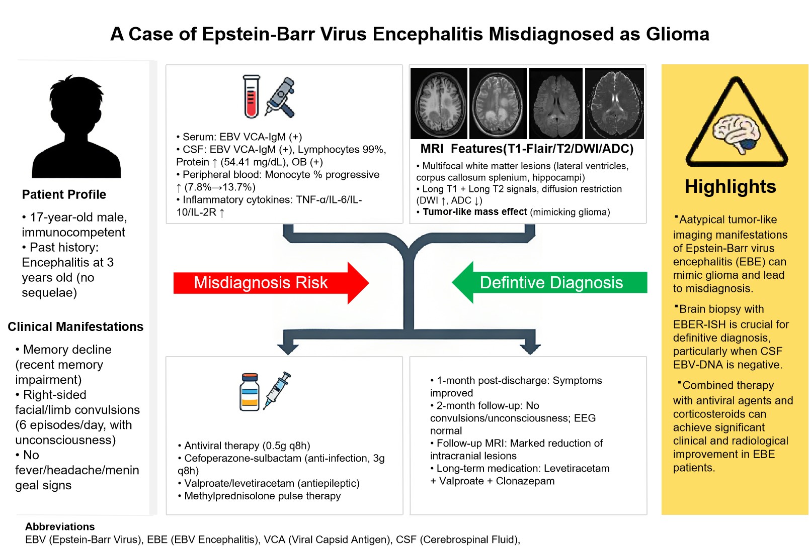
Epstein-Barr virus (EBV) infection can cause central nervous system damage, primarily manifesting as
encephalitis, with diverse imaging
presentations. We report a case of an EBV-infected patient who presented clinically with cognitive
impairment and limb convulsions. Serum
and cerebrospinal fluid tests were positive for EBV viral capsid antigen IgM. Cranial MRI revealed
multifocal white matter lesions
distributed along the lateral ventricles, demonstrating T1 and T2 hyperintensity with associated
diffusion restriction in the involved
areas, patchy enhancement, and a tumor-mimicking appearance with significant mass effect. Brain biopsy
confirmed viral encephalitis. After
treatment with antiviral agents and corticosteroids, the patient's symptoms improved. Follow-up cranial
MRI one month later showed
significant reduction of the lesions. In clinical practice, it is important to be alert to the
tumor-like imaging manifestations of EBV
encephalitis. Pathological biopsy of brain tissue plays a crucial role in distinguishing this condition
from diseases such as glioma.
Keywords: Epstein-Barr Virus Encephalitis; Glioma; Brain biopsy; Tumor-like lesions.
Introduction
Epstein-Barr virus (EBV) is a double-stranded DNA virus that is lymphotropic for humans and belongs to the gammaherpesvirus family (human herpesvirus 4). It is highly prevalent in the human population, with over 90% of adults testing positive for EBV antibodies [1]. Central nervous system damage caused by EBV infection, referred to as Epstein-Barr virus encephalitis (EBE), can manifest as encephalitis, meningitis, transverse myelitis, radiculitis, and Guillain-Barré syndrome (GBS), with meningoencephalitis being the most common presentation [2]. The clinical and imaging features of EBE are highly variable. This article reports a case of EBE presenting with cognitive impairment and tumor-like imaging characteristics, aiming to enhance recognition of the atypical manifestations of this disease.
Case Presentation
A 17-year-old male student was admitted on April 1, 2025, due to memory decline for over two weeks and limb convulsions for 10 days. According to the patient's sister, the patient experienced a rapid decline in memory over two weeks prior without an obvious cause, predominantly affecting recent memory, accompanied by slowed responses. There was no fever, headache, dizziness, nausea, vomiting, loss of consciousness, limb convulsions, or incontinence. A cranial MRI plain scan plus enhancement performed at a local hospital revealed multiple intracranial space-occupying lesions. Ten days prior to admission, the patient experienced episodic right-sided facial and limb convulsions during a hospital visit, accompanied by loss of consciousness, with each episode lasting from 10 seconds to several minutes and occurring up to six times per day. The seizures persisted despite antiepileptic treatment. Since the onset of illness, the patient had poor appetite and sleep, but normal bowel and bladder function, with no significant changes in weight. The family reported a suspected history of acute gastroenteritis prior to the onset of symptoms. Past medical history included encephalitis at the age of three, with no specific details available and no sequelae reported. There were no remarkable findings in personal or family history, and no history of mosquito bites, animal bites/scratches, or exposure to infectious diseases was reported.
Physical Examination: Body temperature 36.2°C, pulse rate 76 bpm, respiratory rate 16 bpm, blood pressure 120/60 mmHg (1 mmHg = 0.133 kPa). The patient was conscious, oriented, but appeared slightly lethargic. Speech was clear, and he was cooperative during the examination. Higher cortical functions including comprehension, orientation, and calculation were grossly intact upon bedside testing. Significant memory impairment was noted, particularly affecting recent memory. Bilateral pupils were equal and round, approximately 3.0 mm in diameter, with normal light reflexes. Cranial nerve examination revealed no deficits. Muscle strength was grade 4 in all four limbs, with normal muscle tone. Deep tendon reflexes were graded 2+ bilaterally. Babinski signs were negative. The neck was supple, and no meningeal signs were observed.
Accessory examination: Complete blood count revealed a progressive increase in the monocyte percentage: 7.8% (reference range 2.0%-11.0%) on 2025-03-23, 11.2% on 2025-04-08, and 13.7% on 2025-04-20. The corresponding absolute monocyte counts were 0.65 × 10⁹/L (reference range 0.14-0.74 × 10⁹/L) on 2025-03-23, 1.11 × 10⁹/L on 2025-04-08, and 1.09 × 10⁹/L on 2025-04-20. Other hematological parameters were within normal limits. Procalcitonin was elevated at 0.093 ng/mL (reference range 0-0.05 ng/mL). Serum inflammatory cytokines showed elevated levels: TNF-α 17.2 pg/mL (reference range 0.0-8.1 pg/mL), IL-6 4.1 pg/mL (reference range 0.0-3.4 pg/mL), IL-10 11.5 pg/mL (reference range 0.0-9.1 pg/mL), and IL-2R 1449 U/mL (reference range 223-710 U/mL). Electrolyte panel indicated hypokalemia (potassium 3.1 mmol/L, reference range 3.50-4.9 mmol/L). Biochemistry tests revealed a blood glucose level of 3.29 mmol/L and elevated homocysteine at 22.7 µmol/L (reference value <15 µmol/L). Serology was positive for Epstein-Barr virus viral capsid antigen IgM (VCA-IgM) and positive for Cytomegalovirus IgG. A respiratory virus panel was positive for Haemophilus influenzae. Routine urinalysis, liver and kidney function, myocardial enzymes, C-reactive protein, erythrocyte sedimentation rate, immune markers and other three items were generally normal.
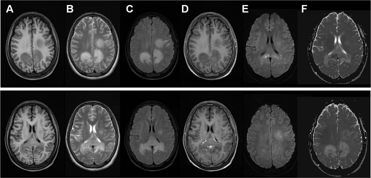
On March 22, 2025, a lumbar puncture was performed for cerebrospinal fluid (CSF) analysis. The CSF appeared colorless and transparent (opening pressure not documented). The white blood cell count was 8 × 10⁶/L (reference range 0-8 × 10⁶/L), with a mononuclear cell predominance of 62.6%. CSF biochemistry showed an elevated protein level of 54.41 mg/dL (reference range 15-45 mg/dL), while glucose and chloride levels were within normal limits. Special CSF protein analysis revealed an elevated CSF albumin level of 0.4 mg/nL (reference range 0.0-0.15 mg/nL). CSF testing was positive for Epstein-Barr virus VCA-IgM. Both serum and CSF oligoclonal bands (OB) were positive. The intrathecal IgG synthesis rate was elevated at 11.32. Serological tests for anti-ganglioside antibodies (AGA), paraneoplastic syndrome-associated antibodies, and autoimmune antibodies were all negative. CSF cytology demonstrated a predominant lymphocytic population (99%). Cranial non-contrast and contrast-enhanced MRI revealed multiple abnormal signal intensities in the splenium of the corpus callosum, fornix, bilateral trigones of the lateral ventricles, bilateral hippocampi, and the left frontal lobe. Patchy enhancement was observed post-contrast administration (Figure 1). Cervical spine MRI showed no significant abnormalities. Following admission, the patient received supportive and symptomatic treatment, including antiviral therapy with acyclovir (0.50g every 8 hours), anti-infective therapy with cefoperazone-sulbactam (3g every 8 hours), and valproate sodium (0.5g twice daily). The patient's symptoms improved, although intermittent right-sided limb convulsions persisted.
Figure 1. Cranial magnetic resonance imaging (MRI) findings on April 2, 2025. A-F show T1-FLAIR, T2, T2-FLAIR, T1-FLAIR+C, DWI, and ADC images, respectively. Irregular T1-hypointense and T2-hyperintense signals are seen in the splenium of the corpus callosum and bilateral centrum semiovale, appearing slightly hyperintense on T2-FLAIR. DWI shows mild hyperintensity with corresponding low ADC values. No significant enhancement is observed post-contrast.
Six days after admission, a brain biopsy was performed. Histopathological examination revealed mild glial hyperplasia, vascular dilation, and perivascular infiltration by numerous lymphocytes and monocytes. Immunohistochemistry results were as follows: H3K27Me3 (partially positive), H3K27M (negative), IDH1R132H (negative), ATRX (positive), P53 (focally positive), BRAF (negative), and Ki-67 proliferation index approximately 5%. Epstein-Barr virus-encoded small RNA in situ hybridization (EBER-ISH) was positive. Based on these collective findings, a diagnosis of EBE was established. Intravenous methylprednisolone pulse therapy (0.25g daily) was initiated. A repeat lumbar puncture four days later showed a CSF pressure of 130 mmH₂O, with both cell count and protein level normalized to within reference ranges. Metagenomic next-generation sequencing of the CSF did not detect any pathogenic microorganisms.
One month after discharge, the patient had largely recovered, with only mild dysarthria and persistent involuntary twitching of the right upper limb remaining. At the two-month follow-up, there were no further episodes of limb convulsions or loss of consciousness. A repeat complete blood count showed a monocyte percentage of 10.7% with an absolute count of 0.64 × 10⁹/L. Follow-up cranial MRI demonstrated a significant reduction in the extent of the previous intracranial abnormal signals. The patient still has involuntary twitching of the right upper limb from time to time. The follow-up electroencephalogram showed normal electroencephalogram. At present, he has been taking levetiracetam, sodium valproate and clonazepam for a long time to control the symptoms.
Discussion
Epstein-Barr virus (EBV) exhibits a high seroprevalence in the general population and can infect individuals of all age groups without distinct seasonal variation. Human is the only host of EBV infection, mainly through the patient's oral saliva, but also through organ transplantation or blood transfusion. EBV infection is associated with a spectrum of conditions, including: (1) primary EBV infection, most commonly presenting as infectious mononucleosis (IM); (2) chronic active EBV infection (CAEBV); (3) EBV-associated hemophagocytic lymphohistiocytosis (EBV-HLH); (4) various malignancies, such as Hodgkin lymphoma (HL), non-Hodgkin lymphoma (NHL), nasopharyngeal carcinoma (NPC), gastric carcinoma, and post-transplant lymphoproliferative disorder (PTLD). Central nervous system involvement by EBV, designated as EBV encephalitis (EBE), is relatively uncommon, affecting approximately 0.4% to 7.5% of infected individuals [3]. Among them, age, underlying immunosuppression, and active peripheral EBV infection have been identified as independent risk factors for the development of EBE [4].
The pathogenesis of EBE remains incompletely understood. The prevailing hypothesis suggests that EBE primarily occurs via retrograde axonal transport of the virus along cranial nerves. However, recent studies have reported the existence of a lymphatic system within the brain that connects the subarachnoid space, venous sinuses, lymphatic vessels, and deep cervical lymph nodes. EBV may potentially exploit this pathway by invading the lymphatic system to gain access to the intracranial compartment, thereby facilitating the development of EBE [5]. Compared to other types of viral encephalitis, meningeal involvement is more frequently observed in EBE patients, implicating the disruption of the blood-brain barrier as a key mechanism in its pathogenesis [4]. Another critical mechanism is the immune response triggered by EBV within the central nervous system. EBV can stimulate B lymphocytes to produce a vast array of antibodies, including those targeting neuronal glycolipid components such as gangliosides. This can lead to widespread parenchymal damage and manifest as various demyelinating diseases, including acute disseminated encephalomyelitis (ADEM), Guillain-Barré syndrome (GBS), and transverse myelitis [6]. Furthermore, studies have reported that reactivation of latent EBV under conditions of immunosuppression contributes to a range of CNS injuries. In the present case, the patient was immunocompetent. Serological and CSF findings were positive for EBV VCA-IgM, while antibodies for VCA-IgG and nuclear antigen (NA)-IgG were negative, and EBV-PCR on the brain biopsy specimen was positive. This profile is consistent with a primary EBV infection, indicating that the virus can directly invade the CNS to cause EBE. However, the patient showed positive cerebrospinal fluid OB test, increased IgG intrathecal synthesis, and multiple tumor-like lesions mainly in white matter on cranial MRI. We speculate that it may be related to immune-mediated neurological damage caused by EBV infection, and a variety of mechanisms were involved in the disease process of this patient.
The clinical manifestations of EBE encompass both systemic and neurological symptoms. Systemic symptoms are typically nonspecific, with headaches, fever, and gastrointestinal issues being the most common. Patients may also experience fatigue, pharyngitis, tonsillitis, myalgia, or urinary disorders. Neurological symptoms predominantly involve impaired consciousness levels and delirium. Other manifestations include cerebellar syndrome (characterized by unsteady gait and ataxia), secondary generalized tonic-clonic seizures, and meningeal irritation signs. Some patients may develop extrapyramidal symptoms or psychiatric disturbances, while cognitive impairment is the primary presentation in rare cases. The patient reported in this case initially presented with rapid memory decline, thereby expanding the known spectrum of EBE symptomatology. Primary EBV infection in immunocompetent individuals is typically asymptomatic or manifests as infectious mononucleosis (IM) [7]. Consistent with previous literature, although this patient did not exhibit significant leukocytosis, a notable increase in the monocyte percentage was observed, which gradually decreased over the course of the illness. However, the correlation between the kinetics of monocyte count and disease progression or prognosis has not been established in the current literature.
The routine cerebrospinal fluid examination results of EBE patients showed no significant difference from other types of viral encephalitis. The white blood cell count was slightly increased, among which 87% of patients showed lymphocyte increase with/without mild protein level increase [8]. The gold standard for EBE diagnosis is the detection of EBV-DNA positive in cerebrospinal fluid or brain biopsy, and the specific EB virus antibody test is the most powerful evidence for the diagnosis of EB virus encephalitis [9]. However, the positive rate of EBV-DNA in cerebrospinal fluid of EBE patients was about 87%, while most of the patients with EBV-DNA negative results were primary EBV infection patients [8]. The CSF analysis of this patient showed a mild increase in lymphocyte-dominated white blood cells with elevated protein levels. However, the EBV-DNA test in the CSF was negative. The possible reasons are as follows: Firstly, the CSF was positive for EBV VCA-IgM while negative for VCA-IgG, indicating a primary EBV infection. Secondly, the lumbar puncture for EBV-DNA testing was performed after the initiation of antiviral therapy; the negative result may therefore be attributed to the timing of the test and a potential decrease in viral load following treatment. It is important to note that the detection of EBV-DNA in the CSF is not entirely specific for EBE, as it has also been associated with central nervous system lymphoproliferative disorders, particularly HIV-associated primary central nervous system lymphoma (PCNSL) [10] and post-transplant lymphoproliferative disorder (PTLD) [11]. This underscores the critical role of brain biopsy in achieving a definitive diagnosis. Previous literature has reported eight confirmed EBE cases where EBV was detected in brain tissue; notably, one of these patients also had a negative CSF EBV-DNA result. In our case, the diagnosis of EBE was ultimately confirmed by a positive Epstein-Barr virus-encoded RNA (EBER) in situ hybridization on brain biopsy tissue. The histopathological findings lacked features typical of glioma, lymphoproliferative disease, or ADEM, thereby solidifying the diagnosis of EBE.
Neuroimaging serves as a crucial diagnostic tool in EBE. Notably, studies indicate that over half of EBE patients show no abnormalities on cranial CT scans, yet abnormal findings emerge on MRI results. This highlights the critical role of cranial MRI in both diagnosis and treatment of EBE [8]. The location of EBE lesions is not significantly specific, and it is more likely to involve the cerebral hemisphere, basal ganglia, cerebellum, brainstem, thalamus and limbic system [12]. The patient's cranial MRI revealed multiple lesions centered around the lateral ventricle, showing T1 low signal, T2 high signal, diffusion restriction, low ADC image intensity, and normal FLAIR image intensity. Mild contrast enhancement was observed (Figure 1). However, some EBE cases exhibit MRI features similar to gliomas and lymphomas, which can easily lead to misdiagnosis in early stages.
The treatment of EBE remains primarily supportive. The Infectious Disease Society of America (IDSA) guidelines recommend empirical antiviral therapy for all suspected encephalitis patients (Grade A recommendation) [13]. However, acyclovir is only a recommended antiviral agent for herpes simplex encephalitis (Class I A). At present, empirical and specific antiviral treatments for EBE are not supported by large randomized controlled trials. When there is immune-mediated demyelinating disease of the nervous system and EBV-associated hemophagocytic syndrome, corticosteroids should be used. In addition to anti-inflammatory and cerebral edema reduction, they can also reduce neurological sequelae. However, the decision to initiate therapy must be guided by a clear clinical rationale, necessitating strict adherence to established indications due to the potential for adverse effects [14].
Abbreviations
ADEM: Acute Disseminated Encephalomyelitis; AGA: An-ti-Ganglioside Antibodies; CAEBV: Chronic Active EBV Infec-tion; CNS: Central Nervous System; CSF: Cerebrospinal Fluid; EBE: Epstein-Barr Virus Encephalitis; EBER-ISH: EBV-encoded RNA In Situ Hybridization; EBV: Epstein-Barr Virus; EBV-HLH: EBV-associated Haemophagocytic Lymphohistiocytosis; GBS: Guillain-Barré Syndrome; HL: Hodgkin Lymphoma; IDSA: Infec-tious Diseases Society of America; IL: Interleukin; IM: Infec-tious Mononucleosis; NA: Nuclear Antigen; NHL: Non-Hodgkin Lymphoma; NPC: Nasopharyngeal Carcinoma; OB: Oligoclonal Bands; PCNSL: Primary Central Nervous System Lymphoma; PTLD: Post-transplant Lymphoproliferative Disorder; VCA: Viral Capsid Antigen.
Declarations
Acknowledgements
None.
Author Contributions
Yinuo Liu collected and analyzed the case data and drafted the initial manuscript. Ru Liu critically revised the manuscript for important intellectual content, including its structure and key statements, and finalized the writing. All authors reviewed and approved the final manuscript.
Funding information
Not Applicable.
Ethics Approval and Consent to Participate
Written informed consent was obtained from the patient for publication of this case report. The procedures were in accordance with the ethical standards of the responsible committee and with the Helsinki Declaration.
Competing Interests
The authors declare that they have no existing or potential commercial or financial relationships that could create a conflict of interest at the time of conducting this study.
Data availability
Not Applicable.
References
[1] Saeed M, Dabbagh O, Al-Muhaizae M, Dhalaan H, Chedrawi A (2014). Acute disseminated encephalomyelitis and thrombocytopenia following Epstein-Barr virus infection. J Coll Physicians Surg Pak, 24 Suppl 3:S216-218.
[9] Tyler KL (2018). Acute Viral Encephalitis. N Engl J Med, 379(6):557–66.
 Figures
 References
 Peer
Peer Information
Figure 1. Cranial magnetic resonance imaging (MRI) findings on April 2, 2025. A-F show T1-FLAIR, T2, T2-FLAIR, T1-FLAIR+C, DWI, and ADC images, respectively. Irregular T1-hypointense and T2-hyperintense signals are seen in the splenium of the corpus callosum and bilateral centrum semiovale, appearing slightly hyperintense on T2-FLAIR. DWI shows mild hyperintensity with corresponding low ADC values. No significant enhancement is observed post-contrast.
[1] Saeed M, Dabbagh O, Al-Muhaizae M, Dhalaan H, Chedrawi A (2014). Acute disseminated encephalomyelitis and thrombocytopenia following Epstein-Barr virus infection. J Coll Physicians Surg Pak, 24 Suppl 3:S216-218.
[9] Tyler KL (2018). Acute Viral Encephalitis. N Engl J Med, 379(6):557–66.
Peer-review Terminology
Identity transparency: Single anonymized
Reviewer interacts with: Editor
Details
This is an open access article under the terms of the Creative Commons Attribution License(http://creativecommons.org/licenses/by/4.0/), which permits use, distribution and reproduction in any medium, provided the original work is properly cited.
Publication History
Received 2025-10-05
Accepted 2025-10-21
Published 2026-03-30